
Research news
Researchers have reported a chip-scale microoptical system that enables continuous, real-time fluorescence monitoring of three-dimensional living microtissues, addressing a longstanding barrier to long-term functional analysis in organ-on-chip models
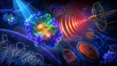
Understanding how living tissue functions over time has remained central to the study of disease mechanisms, evaluation of drug response and the development of advanced in vitro models that more closely reflect human physiology. Researchers have now developed a highly miniaturised microoptical system that has enabled continuous, real-time fluorescence monitoring of three-dimensional microtissues directly on chip-based platforms. By integrating light excitation, signal detection, and tissue confinement within a footprint of approximately one square millimetre, the system has supported prolonged observation of dynamic biological processes without reliance on bulky external optical equipment.
Using fluorescence-based readouts, the platform has captured rhythmic cellular activity as well as rapid responses to stimulation, demonstrating the capacity to track functional changes in living tissues over extended periods with high sensitivity and measurement stability. The approach addresses a persistent technical limitation in microphysiological systems and organ-on-chip technologies, which have seen growing adoption as alternatives to animal experiments and conventional cell cultures.
Despite their promise, these systems have faced challenges in the continuous monitoring of biological activity. Conventional fluorescence imaging has typically relied on large, external microscopes positioned at a distance from the tissue, which has limited scalability, continuous observation and long-term measurement. Repeated handling and transfer steps have also disrupted physiological conditions and introduced experimental variability. Although fluorescence remains a powerful and widely used sensing method, its integration into compact, on-chip platforms has been constrained by the size of optical components and limited system-level integration. These limitations have created a clear need for deeply integrated miniaturised fluorescence monitoring technologies capable of continuous in situ measurement.
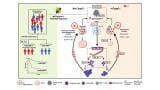
Researchers from KTH Royal Institute of Technology and Karolinska Institutet, both of Stockholm, Sweden, have reported the development of such a system which is a fully integrated microoptical platform designed to enable continuous fluorescence monitoring of living microtissues. The researchers demonstrated that the chip-scale system could integrate directly with microphysiological platforms to track functional activity in three-dimensional tissues over several hours. Validation using pancreatic islet models showed that complex cellular dynamics could be monitored in real time without the use of external optical instrumentation.
The system integrated all essential optical components, including a micro light-emitting diode, a photodetector, optical filters and a custom-designed light guide, into a compact structure measuring approximately 1 mm². A microcage stabilised the tissue mechanically while preserving a physiologically relevant environment. This configuration allowed excitation light to reach the tissue and fluorescence to be observed in close proximity which minimised signal loss and supported stable, long-term measurements directly on chip.
To demonstrate system performance, the researchers monitored pancreatic islets engineered to express a calcium-sensitive fluorescent indicator. Calcium oscillations within these islets have reflected functional activity linked to insulin secretion. The platform successfully recorded rhythmic calcium signals for more than two hours and captured both slow oscillatory behaviour and rapid responses to chemical stimulation. Clear separation between excitation and emission wavelengths reduced background noise and limited phototoxic effects on the tissue.
Compared with conventional fluorescence microscopy, the integrated system has delivered comparable functional readouts while removing the need for bulky optics and manual alignment. Its small footprint has also allowed multiple sensors to operate in parallel, supporting scalable experimental designs and the potential for multi-organ monitoring within interconnected chip platforms.
“This work shows that advanced biological monitoring no longer needs to rely on large external instruments,” said the senior researchers.
“By bringing fluorescence excitation and detection directly next to the tissue, we can observe living systems continuously and with minimal disturbance. This approach makes long-term functional studies more practical and reproducible, particularly for complex microtissues.
“It also creates novel opportunities to integrate sensing technologies into organ-on-chip models that better reflect real physiological conditions,” the added.
The integrated microoptical system offers broad potential for biomedical research and drug development. Continuous in situ monitoring of tissue function could improve studies of chronic disease, drug toxicity and treatment efficacy by enabling detection of slow or subtle biological changes that snapshot experiments often miss.
Beyond pancreatic islets, the platform could support cardiac, neural, or other organoid models and facilitate studies of multi-organ interaction. Its compact and modular design has also suited high-throughput screening and automated analysis, advancing organ-on-chip platforms towards more precise, scalable, and physiologically relevant tools for the study of human health and disease.
For further reading please visit: 10.1038/s41378-025-01073-4
ILM Guide 2026/27


.jpg)


2.jpg)
