Research news
Researchers at the Massachusetts Institute of Technology have developed a compact, low-cost ultrasound system capable of producing wide-angle three-dimensional images, raising the prospect of more frequent screening for people at high risk of developing breast cancer, with use foreseen in community settings and at home
Researchers at the Massachusetts Institute of Technology, Cambridge, Massachusetts, USA, have developed a miniaturised ultrasound system designed to support more frequent breast cancer screening for people at elevated risk, with potential applications in doctors’ surgeries, community clinics or eventually in home settings. The system has combined a small ultrasound probe with a compact acquisition and processing module to allow real-time reconstruction of wide-angle three-dimensional images using a standard laptop computer.
Frequent screening is particularly important for individuals at high risk of breast cancer, as tumours can develop between routine mammograms. These so-called interval cancers account for approximately 20 to 30 per cent of all breast cancer cases and are often more aggressive than tumours detected during scheduled imaging. Early detection remains critical as when breast cancer is diagnosed at the earliest stages, survival rates approach 100 per cent, whereas later-stage diagnoses are associated with survival rates closer to 25 per cent.
The research team reported that the compact design could remove practical barriers that currently limit access to ultrasound imaging, particularly in rural areas and low-resource settings.
“Everything is more compact and that can make it easier to be used in rural areas or for people who may have barriers to this kind of technology,” said Dr. Canan Dagdeviren, associate professor of media arts and sciences at MIT and senior author of the study.
She added that earlier detection of a greater number of tumours could substantially increase the likelihood of successful treatment.
Lead authors were Dr. Colin Marcus and former MIT postdoctoral researcher Dr. Osman Goni Nayeem. Additional contributors included graduate students and research staff from MIT, collaborators from the University of Central Florida, and clinical expertise from a breast cancer surgeon at Massachusetts General Hospital.
Although mammography remains the primary screening tool for breast cancer, ultrasound imaging can provide valuable complementary information, particularly for individuals with dense breast tissue or elevated genetic risk. At present, ultrasound is typically used only as a follow-up investigation after mammography has identified an area of concern. Conventional ultrasound systems are large, expensive and dependent on skilled technicians which has restricted their deployment outside major hospitals.
“You need skilled ultrasound technicians to use those machines which is a major obstacle to getting ultrasound access to [isolated] communities,” said graduate student Shrihari Viswanath, one of the study’s co-authors. The researchers have argued that portability and ease of use are essential if ultrasound is to play a broader role in preventive screening.
The current work has built on earlier research from the same team. In 2023, Dagdeviren and colleagues developed a flexible ultrasound patch that could be attached to a bra and moved across the breast to generate two-dimensional images from multiple angles. Although these images could be combined to form a three-dimensional representation, the approach risked small gaps in coverage and required connection to a large, refrigerator-sized processing unit.
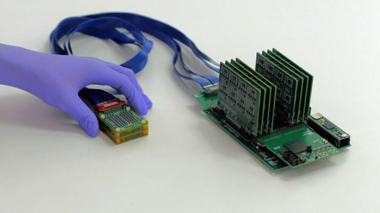
In the latest study, the team aimed to overcome these limitations by developing a fully portable system capable of imaging the entire breast from only two or three positions. The resulting device uses a chirped data acquisition system – dubbed cDAQ – consisting of a probe and a motherboard for data processing. The probe, slightly smaller than a deck of cards, contains an ultrasound array arranged in the shape of an open square, a configuration that enables volumetric imaging of tissue beneath the skin.
The accompanying motherboard is slightly larger than a smartphone and has been built using commercially available electronic components, at an estimated cost of around US$300 dollars. When connected to a laptop computer, the system allows real-time visualisation of three-dimensional ultrasound data, while remaining fully portable.
“A traditional three-dimensional ultrasound system requires expensive and bulky electronics which limit their use to [only within] high-end hospitals and clinics,” said Dr. Anantha Chandrakasan, MIT provost and a co-author of the study.
“By redesigning the system to be ultra-sparse and energy-efficient, this diagnostic tool can move out of the imaging suite and into a wearable form factor that is accessible for patients everywhere,” he added.
The energy requirements of the device are significantly lower than those of conventional ultrasound machines. The system can operate using a five-volt direct current supply, allowing power from a battery or a standard AC to DC adapter commonly used for consumer electronics.
“Ultrasound imaging has long been confined to hospitals. To move ultrasound beyond the hospital setting, we reengineered the entire architecture and introduced a novel ultrasound fabrication process to make the technology both scalable and practical,” said Nayeem.
To evaluate performance, the researchers tested the system on a human volunteer, a 71-year-old woman with a history of breast cysts. The device successfully visualised the cysts and generated a continuous three-dimensional image of the tissue without gaps. The system demonstrated imaging depths of up to 15 centimetres and was able to capture the entire breast from only two or three placements.
Unlike conventional probes, which must be pressed into the tissue and can introduce distortion, this compact device only has to rest gently on the skin.
“With our technology, you simply place it gently on top of the tissue and it can visualise the cysts in their original location and with their original sizes,” Dagdeviren said.
The research team has now initiated a larger clinical study at the MIT Center for Clinical and Translational Research and at Massachusetts General Hospital to further assess performance across a broader population. In parallel, the engineers are working to reduce the size of the processing electronics further, with the aim to create a module approximately the size of a fingernail.
Future versions are intended to connect directly to a smartphone for image visualisation. The team has also planned to develop a smartphone application incorporating an artificial intelligence algorithm to guide users to the optimal probe placement. While the current system could be readily adapted for clinical use, the longer-term goal is to integrate the technology into a wearable sensor suitable for regular home screening by people at high risk of breast cancer.
Dagdeviren has begun work to launch a company to commercialise the technology.
For further reading please visit: 10.1002/adhm.202505310
ILM Guide 2026/27


.jpg)


2.jpg)
