Research news
A research team drawn from Sweden, Germany, Italy and France has created the most complete ‘molecular film’ to date of a self-splicing ribozyme, showing at atomic detail how an RNA catalyst folds, avoids misfolded traps and assembles itself into a functional machine
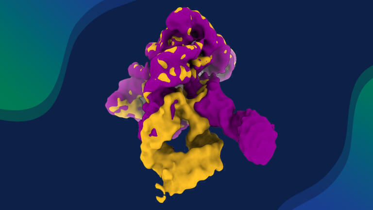
RNA is a central biological macromolecule that underpins gene expression and regulation and underlies many therapeutic modalities and nanotechnology approaches. As with proteins, RNA function depends on a precisely defined three-dimensional structure. A study has now provided what the authors describe as a molecular film of an RNA catalyst – or ribozyme – that assembles itself in real time. The work has visualised how this RNA machine folds, flexes and assembles, and has revealed its structural choreography in unprecedented detail.
The team used an integrative structural biology strategy that combined several state-of-the-art techniques. Cryo-electron microscopy (CEM), small-angle X-ray scattering (SAXS), RNA biochemistry and enzymology, advanced image processing and molecular simulations all contributed complementary information. Together, these methods allowed the researchers to follow the assembly of a self-splicing ribozyme, an RNA molecule that can cut and rejoin its own sequence to generate a functional product without the need for protein enzymes. In effect, the molecule edits itself to become operational.
The study, led by the group of Dr. Marco Marcia, former group leader at the European Molecular Biology Laboratory (EMBL) in Grenoble and now Associate Professor and Science for Life Laboratory group leader at Uppsala University in Sweden, has captured the dynamic route by which a self-splicing ribozyme folds into its active structure. By reconstructing successive structural states, the researchers have moved beyond static pictures and provided a mechanistic account of RNA folding in action.
The work has relied on advanced infrastructure at EMBL Grenoble, which enabled the integration of high-end structural biology methods with RNA biochemistry and enzymology. The Marcia group also collaborated closely with the Centre for Structural Systems Biology (CSSB) in Hamburg, where project-specific CEM image-processing approaches were developed, and with the Italian Institute of Technology in Genoa, which provided expertise in molecular simulations at high resolution.
“Determining RNA structures is a challenging task – the inherent flexibility and negative charge make RNA a notoriously difficult target for structural studies. Persistent efforts and extensive screening on electron microscopes ultimately led us to visualise elusive RNA dynamics.” said Dr. Shekhar Jadhav, formerly of EMBL Grenoble but now a postdoctoral researcher at Uppsala University.
That combination of persistence and technical refinement has produced the most complete molecular film so far of an RNA molecule that builds itself.
The resulting dataset shows how the ribozyme avoids the biological equivalent of cinematic outtakes: misfolded, non-functional conformations known as kinetic traps. Rather than collapse into dead-end structures, the ribozyme follows a sequence of productive states that guide it towards the configuration required for catalysis. The work therefore addresses a fundamental question in RNA biology, namely how large RNA molecules navigate a complex energy landscape to reach a native, functional fold.
Central to this process is Domain 1 (D1), the core scaffold of the ribozyme. The study portrays D1 as both structural framework and director of the unfolding scene. This domain acts as a molecular gate that cues other domains – designated D2, D3 and D4 – to dock at specific stages of the folding trajectory. Subtle structural shifts in key regions of D1 cause one part of the domain to open and create the correct environment for the next domain to engage.
Each additional domain only joins once the previous elements have occupied their appropriate positions. The result is a tightly ordered sequence of molecular events that minimises structural error and culminates in a correctly formed active site capable of catalysis.
To reach this level of detail, the researchers analysed hundreds of thousands of individual ribozyme particles. From this large ensemble they reconstructed intermediate ‘takes’ that remain invisible to more traditional crystallographic methods, which tend to average over many molecules and focus on stable end states. The intermediate frames show how the RNA samples alternative arrangements before it settles into its final conformation.
“To capture these fleeting frames, we had to develop novel CEM image-processing strategies,” said Professor Maya Topf, group leader at CSSB at the University Medical Centre Hamburg-Eppendorf, and a collaborator on the study.
“This is a great example of how computational innovation and high-quality CEM data can reveal the hidden conformations of molecular machines,” she said.

By tailoring the image-processing pipeline to this problem, the team extracted subtle structural differences from noisy data, which allowed consecutive stages of the folding pathway to emerge.
SAXS measurements and molecular dynamics simulations provided orthogonal evidence that supported and extended the CEM findings. SAXS contributed low-resolution information about overall particle shapes in solution, while the simulations explored how the molecule moved within the constraints defined by the experimental structures. Together, these methods revealed a high degree of conformational plasticity and allowed the scientists to refine each structural frame and assemble a coherent narrative of the folding process.
The calculated energy barriers between different conformational states appeared relatively small. That feature helps explain how the ribozyme can move smoothly between shapes in the cell and also means that computer simulations can more faithfully reproduce these transitions without the molecule becoming trapped in unrealistic configurations.
“One major strength of this work is the synergy between these cutting-edge novel structural data on RNA and our advanced molecular simulations of this challenging system,” said Dr. Marco De Vivo, head of the molecular modelling and drug discovery laboratory and associate director for computation at the Istituto Italiano di Tecnologia in Genoa, and a co-author of the study.
“This combined approach has clarified, at an unprecedented atomistic level of detail, the dynamic that drives the entire assembly of this RNA molecule, which now opens new avenues for drug discovery efforts targeting RNA,” he added.
The detailed view of how the ribozyme assembles could, for example, highlight vulnerable states that small molecules or antisense agents might stabilise or disrupt. The ribozymes that feature in this work belong to the group II introns, a family of self-splicing elements that many researchers regard as evolutionary precursors of the spliceosome, the large ribonucleoprotein machine that edits precursor messenger RNA in modern eukaryotic cells.
By clarifying how group II introns fold efficiently and avoid kinetic traps, the study offers insight into how early RNA-based life may have evolved the first RNA editing tools. The findings strengthen the long-standing hypothesis that contemporary splicing factors and spliceosomal RNAs have their roots in ancestral ribozymes with self-splicing capacity.
Beyond evolutionary questions, the work provides a template for rational RNA design. A detailed map of productive and non-productive folding trajectories can guide efforts to script RNA molecules that fold reliably for use in therapeutics or nanobiotechnology. For example, messenger RNA-based vaccines, RNA aptamers or RNA components in nanoscale assemblies could all benefit from design principles that avoid kinetic traps and favour robust attainment of the desired native fold.
The extensive datasets and mechanistic insights also represent a valuable benchmark for artificial intelligence. The authors noted that some of the RNA structures solved in the study have already contributed to international Critical Assessment of Structure Prediction exercises, the community-wide challenges that have driven progress in protein and nucleic acid structure prediction and that were instrumental in the rise of AlphaFold, the Google Deepmind-developed AI system that predicts protein 3D structures from amino acid sequences.
“This work is expected to play a key role in shaping artificial intelligence approaches to RNA structure prediction, paving the way towards a new ‘AlphaFold for RNA’,” said Marcia. In this context, the combination of experimentally constrained structural intermediates and physically grounded simulations provides the type of reference dataset that machine-learning methods require if they are to capture not only static structures but also the dynamic behaviour of RNA.
This convergence of experimental precision and machine learning signals a significant phase for RNA structural biology. CEM, SAXS, biochemical assays and complementary experimental approaches now provide rich, time-resolved information on molecular motions, while artificial intelligence methods learn to predict those motions from sequence alone. As the two strands inform each other, researchers expect to predict, visualise and understand in far greater depth the dynamics of one of life’s most versatile molecules.
For further reading please visit: 10.1038/s41467-025-65502-8
ILM Guide 2026/27


.jpg)


2.jpg)
