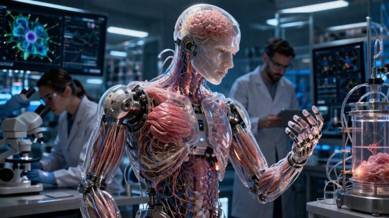
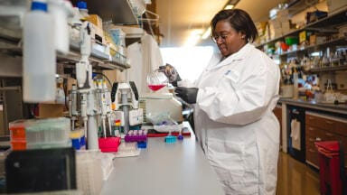
![[From left] Xavier Trepat, Pau Guillamat and Marino Arroyo. Xavier Trepat is an ICREA Research Professor at IBEC, principal investigator of the Integrative Cell and Tissue Dynamics group, and co-lead author of the study. Pau Guillamat is a postdoctoral researcher in the Integrative Cell and Tissue Dynamics group at IBEC and the study's first author. Marino Arroyo is full professor in the Department of Civil and Environmental Engineering at the Polytechnic University of Catalonia (UPC), and co-lead author of the study. Credit: Institute for Bioengineering of Catalonia (IBEC)](/assets/file_store/pr_files/67320/thumbnails/images/768w-432h-t-fit-trepat-guillamat-arroyo-programmable-cell-orientation-living-tissue.jpg)
Research news
Researchers in Barcelona have demonstrated that chemical patterning can direct cell orientation within tissues which enables controlled force generation and predictable three-dimensional shaping with implications for tissue engineering and biohybrid robotics
Biological tissues have long exhibited a striking capacity to organise and to adopt complex shapes through forces generated intrinsically by their constituent cells. To harness this behaviour has remained a central challenge in bioengineering, where the ambition has been to design synthetic living materials capable of reproducible and predetermined morphologies.
A study led by Institute for Bioengineering of Catalonia (IBEC), Polytechnic University of Catalonia (UPC) – BarcelonaTech and International Centre for Numerical Methods in Engineering (CIMNE), all in Barcelona, Spain, and in collaboration with European Molecular Biology Laboratory, which is distributed across Europe but headquartered in Heidelberg, Germany. has now reported a strategy to programme such shape changes through controlled cell orientation using chemical patterning.
The researchers have shown that tissues can deform in a controlled and reproducible manner to generate defined three-dimensional structures. This result has addressed a longstanding obstacle in the field, namely to direct internal cellular forces with sufficient precision to dictate final tissue geometry. The work has focused on the manipulation of how cells align within a tissue which in turn governs the mechanical stresses that drive deformation.
“We are demonstrating that we can design the shape [that] a living tissue will adopt just by controlling how its cells are oriented,” said Dr. Xavier Trepat, a Catalan Institution for Research and Advanced Studies (ICREA) research professor at IBEC, who co-led the study. He has also held positions at the University of Barcelona and within the Centre for Biomedical Research Network in Bioengineering, Biomaterials and Nanomedicine.
At the core of the approach lies the concept of ‘nematic order’ – a physical principle more commonly associated with liquid crystals. In biological tissues composed of elongated cells, this order has emerged as cells align collectively in a shared direction, analogous to fibres within a textile. Such alignment has not been uniform, however, and can break down at discrete points known as topological defects. These defects have represented local disruptions in order and have acted as focal points for force generation, with consequences for tissue growth, migration and deformation.
“The orientation of the cells controls the forces, and the forces can control the generation of a three-dimensional shape,” said Dr. Pau Guillamat, first author of the study. This relationship between orientation and force has formed the theoretical and experimental basis of the work.
To impose control over these processes, the team has used chemical micropatterning techniques to define adhesion landscapes on flat substrates. Lines of adhesive protein have guided cell alignment, while surrounding regions coated with non-adhesive polymer have prevented attachment. This approach has enabled the researchers to construct precise maps of cellular orientation across the tissue. By design, these maps have dictated the spatial placement of topological defects which in natural systems arise spontaneously and without order.
“The key is that we can decide where these defects will be and therefore where the forces will be generated within the tissue,” Guillamat explained. This capacity to localise force generation has provided a route to programme tissue deformation with spatial accuracy.
A critical phase of the experiments has involved the mechanical release of the tissue from its substrate. While attached, the forces generated by cellular alignment have remained constrained. Upon detachment, these stresses have redistributed and the tissue has undergone rapid contraction and deformation in accordance with the pre-imposed orientation patterns.
“It’s like an elastic sheet that is stretched tight and fixed at the edges. While it is held in place, it does not deform. However, when released, it adopts a geometry determined by the internal stresses,” Guillamat said.
This analogy has captured the essential physics of the system, in which stored mechanical energy drives morphological transformation once constraints are removed.
To complement the experimental work, theoretical modelling has been developed under the leadership of Dr. Marino Arroyo at UPC and CIMNE. These models have enabled the prediction of how specific orientation patterns translate into three-dimensional forms. By integrating simulation with experimental data, the team has established a quantitative framework that links nematic order to final tissue shape.
“Our models have allowed us to examine different hypotheses and ultimately identify the mechanism by which cell orientation leads to the three-dimensional folding of tissues.
“Furthermore, they provide a quantitative relationship between the nematic pattern and the shape,” Arroyo said.
This predictive capability has positioned the system as a platform for rational tissue design rather than empirical trial. The implications of this work have extended beyond proof of concept. The ability to programme living tissues without reliance on artificial scaffolds has presented novel opportunities in tissue engineering, where structural fidelity and biological compatibility are essential.
In parallel, the findings have suggested routes to develop biohybrid robotic systems in which living tissues function as actuators, capable of controlled deformation and response. The concept of ‘smart’ living materials has also emerged, with tissues able not only to adopt specific shapes but potentially to respond dynamically to environmental cues.
“These systems can be considered living materials that generate programmable forces and shapes and can also integrate information and respond intelligently,” Guillamat added.
Beyond application, the methodology has provided a powerful experimental framework to investigate fundamental biological processes. By enabling precise manipulation of cell orientation and force distribution, the approach has offered insight into phenomena such as organ development and tumour progression, where mechanical cues play a decisive role.
“It is a perfect tool to understand how patterns of cell orientation influence the mechanics and evolution of complex tissues,” Trepat said.
For further reading please visit: 10.1126/science.adz9174
ILM Guide 2026/27


2.jpg)
