Research news
A research team drawn from collaborators across the USA has developed a super-resolution photoacoustic imaging method that tracks individual red blood cells in the brain, offering a closer view of how microvessels redirect blood flow and oxygen after stroke and potentially opening a route to better understanding of vascular dementia, Alzheimer’s and cerebral small vessel disease
The brain depends on a constant, precisely regulated supply of oxygen and nutrients, delivered through an intricate network of microvessels that extends through neural tissue. Although modern brain imaging has allowed scientists to monitor the activity of individual neurones with remarkable precision, the ability to examine the brain’s smallest blood vessels has remained limited. That technical shortfall has restricted efforts to understand cerebral small vessel disease.
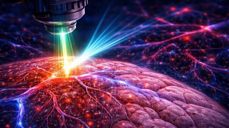
Here, researchers at Washington University (WashU) in St. Louis and Northwestern University have developed a microscopy platform designed to close that gap. Led by Professor Song Hu, of the department of biomedical engineering in the McKelvey School of Engineering at Washington University in St. Louis, USA, the team has created a super-resolution functional photoacoustic microscopy (SR-fPAM).
The method has allowed the researchers to track the movement of individual red blood cells and measure oxygenation-linked changes in their optical properties, producing images of blood flow and oxygen distribution in mouse model brains at the resolution of single-cells.
The study described a way to visualise microvascular function with far greater detail than conventional photoacoustic approaches. The advance is important because the brain’s smallest vessels play a central part in tissue survival, metabolic support and neuronal performance, yet they remain difficult to examine in living tissue with both structural and functional detail at once.
Photoacoustic microscopy relies on a well-established physical principle. Haemoglobin in red blood cells absorbs light naturally because it carries oxygen. When exposed to short laser pulses, that absorbed energy generates ultrasound waves, a process known as the ‘photoacoustic effect’. Scientists can detect those waves to build images of blood vessels without the need to add fluorescent or radioactive labels. Conventional photoacoustic microscopy has already proved useful for vascular imaging, but it has not offered true three-dimensional single-cell resolution.
The team sought to overcome that limitation by building a high-speed photoacoustic microscope capable of image capture from the same brain region at millisecond intervals. That speed allowed the researchers to follow red blood cells as they moved in single file through capillaries and in larger groups through wider vessels. By track these cells from frame to frame and combine their trajectories through computational reconstruction, the researchers produced three-dimensional maps of the brain’s microvascular network at single-cell resolution.
“Similar to super-resolution fluorescence and ultrasound imaging, SR-fPAM leverages high-speed imaging to track dynamics and uses that information to identify features that are smaller than the conventional resolution limit,” Professor Hu said.
“We condense multiple spatiotemporally acquired frames into a single one with substantially improved resolution,” he said.
That principle places SR-fPAM within a broader family of super-resolution methods that use movement over time to infer details finer than the nominal limit of the instrument itself. In this case, the moving signal source is the red blood cell which serves not only as a passive marker of vessel structure but also as a functional reporter of oxygen transport. That combination gives the method unusual power as it can show where blood travels, how fast it moves and how oxygen delivery changes within the same three-dimensional microvascular landscape.
In experiments designed to test the technique under pathophysiological conditions, the researchers used SR-fPAM to examine what happened after an induced stroke in the brain of a mouse. They found that when a single microvessel became blocked, neighbouring vessels altered their flow patterns almost immediately. Red blood cells diverted through alternative routes, which helped preserve oxygen supply to tissue placed at risk by the obstruction. Those observations offered a direct view of the brain’s capacity to compensate for local vascular injury at the smallest scale.
“When one vessel is blocked, red blood cells take alternative routes to continue the flow and oxygen supply,” Professor Hu said.
“Using SR-fPAM, we can observe not only structural changes in the 3D microvasculature but also how fast red blood cells move, how their flow directions change, and how they release oxygen into the surrounding tissue in response to stroke-induced ischemia,” he added.
That finding matters because cerebral injury often begins at the microvascular level long before gross tissue loss becomes obvious. In stroke, vascular dementia and Alzheimer’s disease, disruption to local blood flow and oxygen handling can contribute to neuronal stress, tissue dysfunction and progressive cognitive decline. A method that can reveal such changes at single-cell resolution could therefore help researchers identify early disease mechanisms that remain hidden with current imaging tools.
The authors said how they want to combine SR-fPAM with two-photon microscopy, an imaging technique widely used to visualise neurones and cellular signalling in living tissue. Pairing these techniques could allow simultaneous observation of neuronal activity and microvascular behaviour, both at single-cell resolution, within the same region of brain tissue.
“This would allow us to study how neurons and microvessels are spatiotemporally coordinated with each other and how their dynamic coupling gets disrupted in disease.
“It may also help us better interpret clinical neuroimaging techniques, such as functional magnetic resonance imaging, which infers brain activity from vascular signals,” Professor Hu said.
That prospect is especially significant because functional MRI, does not measure neuronal firing directly. Instead, it infers brain activity from changes in blood flow, blood volume and oxygenation. A tool such as SR-fPAM could therefore help researchers test, refine and interpret the vascular assumptions that underpin widely used neuroimaging methods. In turn, that could strengthen the biological interpretation of clinical scans and improve confidence in studies of brain function and disease.
Professor Hu said the work could have notable translational value as the field seeks better ways to detect and treat vascular contributions to dementia.
“Cerebral small vessel disease is increasingly recognized as a leading cause of cognitive impairment and dementia, and WashU is at the frontier of this in both basic and clinical research,” Professor Hu said.
“If we can better understand how microvascular oxygenation and flow change in the early stages of disease, it may help guide the development of early detection strategies and therapeutic interventions.”
Taken together, the findings have presented SR-fPAM as more than a technical refinement. The method offers a way to observe the living brain’s smallest vascular responses in real time and in three dimensions, with individual red blood cells as the unit of measurement.
For researchers who study how vascular dysfunction intersects with neurodegeneration, stroke and cognitive decline, that is a meaningful step forward. It has moved the field closer to a view of the brain in which blood supply and neural function can be studied side by side.
For further reading please visit: 10.1038/s41377-026-02235-3
ILM Guide 2026/27


.jpg)


2.jpg)
