Research news
Cryo-electron tomography has revealed how flaviviruses reorganise infected cells to enable replication and maturation with implications for future tick-borne encephalitis treatments
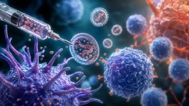
Researchers at Umeå University, Sweden, have shown how tick-borne viruses reorganise human cells to create highly specialised environments that support viral replication and maturation. The findings have provided detailed structural insight into how these pathogens propagate within host cells and may inform future strategies to treat tick-borne encephalitis.
Tick-borne encephalitis is one of the most serious viral infections transmitted in Europe. The disease arises following a bite from an infected tick and can lead to severe inflammation of the brain, with potentially long-lasting neurological consequences. Despite its clinical importance, direct investigation of the causative virus has remained constrained by biosafety limitations.
To overcome this barrier, the research team has used Langat virus, a closely related but less pathogenic member of the flavivirus genus. This surrogate system has allowed researchers to examine intracellular viral processes in detail while maintaining experimental safety.
“When we saw the three-dimensional images for the first time, we immediately realised how much new information we could gain about the virus’ replication,” said Dr. Lars Anders Carlson, professor at the Department of Medical Chemistry and Biophysics at Umeå University, who led the study.
The study has relied on cryo-electron tomography, an advanced imaging approach that enables visualisation of cellular structures in near-native conditions. By rapidly freezing infected cells, the method has preserved delicate viral and cellular architectures which has allowed researchers to reconstruct high-resolution, three-dimensional images of the intracellular environment.
These reconstructions have shown that infection induces extensive remodelling of internal cell membranes. The virus creates discrete compartments that function as protected ‘gene factories’, within which viral RNA synthesis occurs. This organisation appears to shield replication intermediates from host defences while enabling efficient production of viral components.
The study has also clarified the spatial relationship between replication and particle assembly. Newly formed viral particles have emerged in close proximity to these replication compartments, suggesting a tightly coordinated process that minimises transport requirements within the cell. Structural analysis has demonstrated how these particles transition from immature to mature forms prior to release, a critical step in the viral life cycle that determines infectivity.
By comparing two closely related viral variants, the researchers have identified how minimal genetic differences can influence this maturation process. Even a single nucleotide change has altered the rate at which viral particles acquire their mature, infectious configuration.
“Here we were able to directly observe how a small change in a single gene caused the virus to mature at different rates,” said Dr. Bina Singh, postdoctoral researcher on the team.
This observation has underlined the sensitivity of viral replication dynamics to minor genetic variation, a factor that may contribute to differences in virulence, transmission efficiency and response to host immunity. Such insights are particularly relevant for flaviviruses, a group that includes several medically important pathogens.
The work has also highlighted the technical and organisational demands required to achieve this level of structural resolution. Cryo-electron tomography of infected cells and tissue samples requires not only specialised instrumentation but also highly coordinated experimental design, sample preparation and computational analysis.
The project originated within the Umeå Centre for Microbial Research which has supported interdisciplinary collaboration across infection biology. Early-stage funding has enabled the recruitment of international researchers, including Dr. Jianguo Zhang and Dr. Erin Schexnaydre, through the centre’s Excellence by Choice programme.
Within the laboratories of Carlson and Dr. Anna Överby, and in collaboration with the Umeå Centre for Electron Microscopy, these researchers have developed and refined methodologies to visualise tick-borne virus infection in both cultured human cells and mouse brain tissue. This dual approach has strengthened the biological relevance of the findings by linking cellular observations to tissue-level infection contexts.
The study has subsequently expanded into an international collaboration that has involved partners in Norway and the USA. Financial support from the Swedish Research Council and the Knut and Alice Wallenberg Foundation has enabled the integration of complementary expertise and resources.
Taken together, the findings have provided a detailed account of how tick-borne flaviviruses exploit host cell architecture to sustain replication. By clarifying the structural basis of viral assembly and maturation, the work has established a platform for future studies to target these processes therapeutically.
For further reading please visit: 10.1038/s41467-026-68483-4
ILM Guide 2026/27


2.jpg)
