Research news
Currently, there is no way to reverse spinal cord injuries
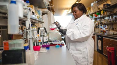
For the first time, researchers at the University of Minnesota Twin Cities, Minneapolis and Saint Paul, Minnesota, have demonstrated a process that combines three-dimensional (3D) printing, stem cell biology and laboratory-grown tissues to promote recovery after spinal cord injury.
According to the United States’ National Spinal Cord Injury Statistical Center, more than 300,000 Americans live with spinal cord injuries. There has so far been no way to fully reverse the spinal cord damage or its resulting paralysis. A major obstacle has been the death of nerve cells and the failure of nerve fibres to regenerate across the injury site. The Minnesota team has now reported a method to address this challenge.
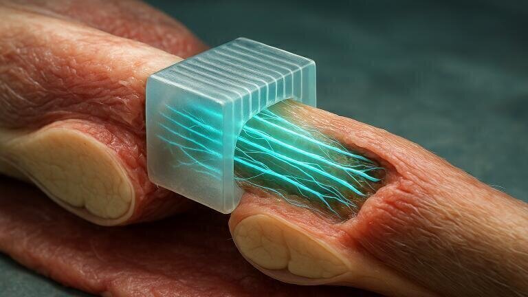
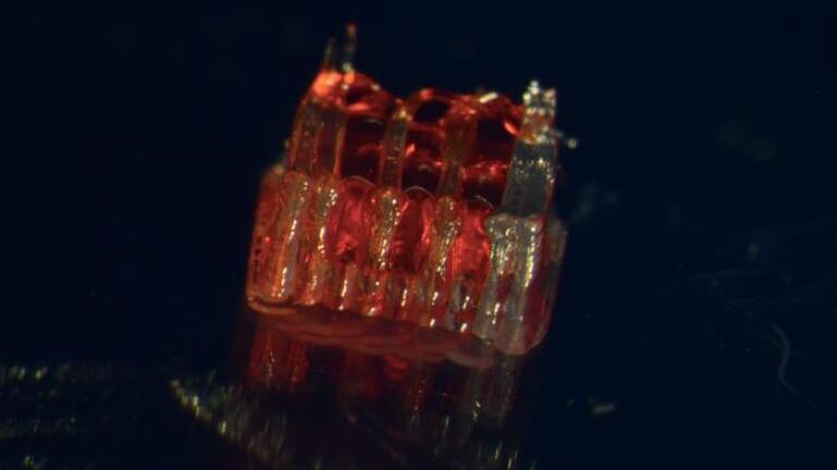
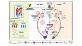
The approach involves the design of a 3D printed framework for laboratory-grown organs – termed an organoid scaffold – with microscopic channels. These channels were populated with regionally specific spinal neural progenitor cells, derived from human adult stem cells which are able to divide and differentiate into distinct types of mature cells.
“We use the 3D printed channels of the scaffold to direct the growth of the stem cells, which ensures the new nerve fibres grow in the desired way,” said Dr. Guebum Han, a former University of Minnesota postdoctoral researcher in mechanical engineering and first author of the study, who now works at Intel Corporation.
“This method creates a relay system that when placed in the spinal cord bypasses the damaged area,” he added.
In their experiments, the team transplanted the scaffolds into rats whose spinal cords had been completely severed. The cells successfully differentiated into neurons and extended fibres rostrally towards the head and caudally towards the tail, forming new connections with the animals’ existing nerve circuits. Over time, the transplanted cells integrated into the host spinal cord tissue and enabled significant functional recovery.
“Regenerative medicine has brought about a novel era in spinal cord injury research,” said Dr. Ann Parr, professor of neurosurgery at the University of Minnesota.
“Our laboratory is excited to explore the future potential of our ‘mini spinal cords’ for clinical translation,” she said.
Although still at an early stage, the research has offered a promising avenue for spinal cord repair. The scientists have expressed the intention to scale up production and further refine the technology with the aim of eventual clinical application.
For further reading please visit: 10.1002/adhm.202404817
ILM Guide 2026/27


.jpg)


2.jpg)
