Research news
Researchers at the University of Texas at Dallas have developed a biosensor that, when integrated with artificial intelligence, can identify volatile organic compounds in exhaled breath linked to thoracic cancers. The technology could pave the way for affordable, non-invasive screening in primary care
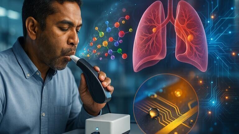
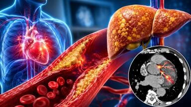
Researchers at the University of Texas at Dallas have developed a biosensor that – in combination with artificial intelligence (AI) – detects volatile organic compounds (VOCs) in breath exhalate that are associated with thoracic cancers. The electrochemical device can identify eight VOCs that may serve as biomarkers for lung and oesophageal cancers. AI then analyses their biochemical signatures to determine whether they correspond to profiles linked to those malignancies.
“We built a screening tool that could allow physicians to catch the disease in its early phases, which improves outcomes,” said Professor Shalini Prasad, head of bioengineering in the Erik Jonsson School of Engineering and Computer Science.
“This technology offers a potentially affordable, quick and non-invasive breath-analysis tool for cancer screening,” she added.
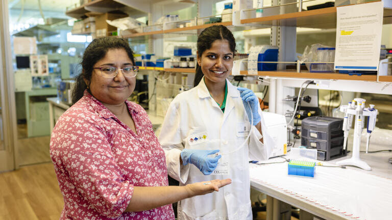
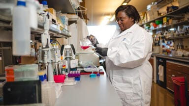
The project united bioengineering and computer-science specialists from the university with a clinical research team from the University of Texas Southwestern Medical Center, Dallas.
In testing, the biosensor was used to analyse breath samples from 67 participants, including 30 whose thoracic cancers had been confirmed by biopsy. The biosensor correctly identified the volatile compounds in 90 per cent of the confirmed cases.
Professor Prasad explained that the inspiration for the project emerged during the COVID-19 pandemic, when non-invasive diagnostic technologies became an urgent focus of research.
“The use of breath became very attractive because breath goes through our respiratory system and carries metabolites, which are indicators of disease,” she said.
Changes in metabolites within exhaled breath may occur early in the onset of disease. This growing field of study – known as breathomics – has the potential to allow clinicians to assess VOCs as diagnostic or prognostic indicators. Professor Prasad emphasised that AI is a vital component of the diagnostic potential of the device.
“There is a huge amount of data provided by the breath.
“What is important? What is not? All of this information comes from the machine-learning algorithm.
“That is why the partnership with computer science is critical. How meaningfully you integrate AI into a technology is important,” she said.
Professor Prasad collaborated with Professor Ovidiu Daescu, head of computer science and the Jonsson School Chair, to refine and validate the machine-learning models.
“The breath-profiling device and associated machine-learning model have great potential for making a difference in cancer detection while improving costs, assuming more cases are tested and validated over time in medical settings,” Professor Daescu said.
Further clinical insight was provided by Professor of Internal Medicine in the Division of Pulmonary and Critical Care Medicine at UT Southwestern, Dr. Muhanned Abu-Hijleh, who served as medical director of respiratory therapy and director of the chronic obstructive pulmonary disease programme.
“Lung cancer is the leading cause of cancer-related deaths in the United States and worldwide.
“Using minimally invasive technologies like biomarkers and exhaled volatile-organic-compound analysis can help in the early detection of thoracic malignancies with minimal burden on patients and the health care system, carrying less overall morbidity,” he said.
Professor Prasad said that the team intends to continue refining the biosensor and to undertake further clinical validation.
“Eventually, this technology could be deployable in your primary-care provider’s office.
“So just as you go in for an annual physical and give an annual blood draw, you could do a breath test as well. Then the primary-care provider could make recommendations if the indicators are elevated, such as a follow-up referral,” she said.
If validated in larger and more diverse patient cohorts, the technology could mark a significant advance towards accessible, cost-effective screening for thoracic cancers, reducing both diagnostic delays and the physical burden on patients.
The study’s contributors included Dr Anirban Paul, a research scientist in bioengineering; Kordel France, a doctoral candidate in computer science; and Avi Bhatia, a senior neuroscience student in the School of Behavioural and Brain Sciences. Collaborators from UT Southwestern included Ruby Thapa, clinical research assistant, and Dr Rhoda Annoh Gordon, research programmes manager in pulmonary and critical care medicine.
For further reading please visit: 10.1016/j.sbsr.2025.100815
ILM Guide 2026/27


.jpg)


2.jpg)
