Microscopy & microtechniques
Published over 15 years ago. See the latest and most current information on Microscopy & microtechniques.
… then have a look what Bruker has to offer you in the area of microstructural analysis:
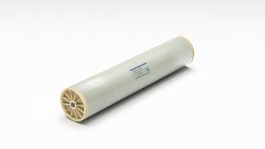
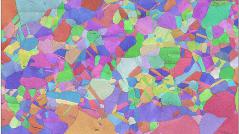
Electron Backscatter Diffraction (EBSD) is the not so new companion to X-ray diffraction (XRD) as a method for the analysis of the microstructure of crystalline samples. Although it requires an effort in sample preparation and – of course – some investment in instrumentation, the effort and the expense are well worthwhile. Compared to XRD, EBSD provides you with spatially resolved information. You will therefore be able to make connections between the material and effects of it’s processing in dependence on the analysed location. This is – for instance – very valuable when looking at heat-treated metals.
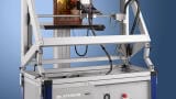
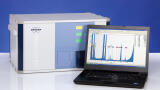
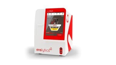
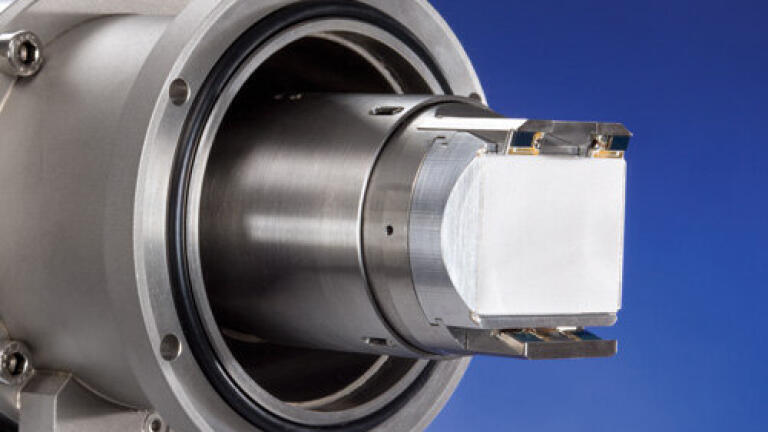
What you need is a scanning electron microscope (preferably with field emission gun) and an EBSD analysis system like Bruker’s revolutionary QUANTAX CrystAlign. CrystAlign consists of the unique e-Flash1000 EBSD detector, the ESPRIT EBSD software and a high-speed computer workstation. The e-Flash1000 records so-called EBSD patterns generated during the scan of the electron microscope’s electron beam across the sample surface and streams them to the workstation. These patterns contain the sought after information on the crystalline structure of the sample. Evaluated and assembled to different representations by the ESPRIT EBSD software, they allow drawing of many conclusions on the nature of the sample. First of all crystalline phases can be identified, then the orientations of crystallites with respect to different reference systems can be analyzed, grains and their size can be determined, and – as advanced options – information like local stress and deformation can be analysed. This just to name some of the many possibilities EBSD analysis offers.
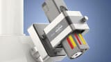
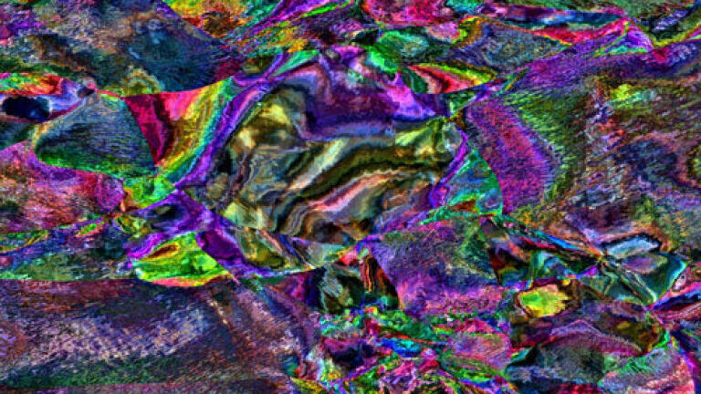
Recently, Bruker has added the new ARGUS™ system of forescattered / backscattered electron (FSE/BSE) detectors as accessories to the e-Flash1000. The BSE detectors provide the usual density-dependent signal known from the BSE detectors that come with the microscope, only that these might not deliver a good signal due to the high sample tilt angle required for EBSD measurements. This is where the ARGUS™ BSE detectors come handy for general sample inspection and positioning. The FSE detectors – in comparison – see the same structure dependent information that the EBSD detector sees. This signal is highly anisotropic, so that the three FSE detectors arranged in a row below the EBSD detector screen see different signal intensities. The intensities will vary with changes in the sample microstructure. So colour-coding and mixing the signals of the three detectors will not only produce extremely colourful images but will also provide a fast qualitative overview of the sample microstructure. This is e.g. very useful for judging sample preparation quality and finding the locations suitable for the more time consuming EBSD analysis.
ILM Guide 2026/27