Microscopy & microtechniques
Published over 14 years ago. See the latest and most current information on Microscopy & microtechniques.
Scientists at the Department of Biochemistry, the University of Oxford, rely on a number of powerful imaging systems, several of which are supplied by Preston based Image Solutions (UK) Ltd. Their efforts are directed to understand how cells become polarised during embryonic development.
Defects in this polarisation process are known to cause birth defects, and also are similar to the processes that go wrong in Alzheimer’s disease and Fragile X Syndrome. By studying neurons of the fruit fly Drosophila, they are trying to understand how RNA, a molecule related to DNA, moves and becomes localised during this process.
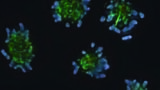
“We know that RNA has a role in its own right and one that can be localised in the cytoplasm a long way from the cell nucleus. A very good example of this occurs in Drosophila neurons, where the RNA can be localised at the cell extremities, where it is regulated and controlled locally. This local control and regulation is known to be important in human diseases such as Alzheimer’s and Fragile X Syndrome,” explained Professor Ilan Davis, Welcome Trust Senior Research Fellow with the Department of Biochemistry.
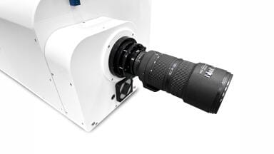
Professor Davis and his team use three DeltaVision Core systems and an OMX ‘super resolution’ instrument to study how RNA behaves in real time, in living cells. The DeltaVision Core is designed as an imaging workhorse to image a large number of probes and samples with great precision. The OMX is a more specialised and advanced instrument that uses three dimentional structured illumination technology, developed by a team at the University of California San Francisco (UCSF): this doubles the spatial and axia resolution of a widefield light microscope. Additionally, this system can deliver high temporal resolution data for the study of fast dynamics in widefield mode. One DeltaVision Core has been modified to work with an upright Olympus platform microscope, rather than the usual inverted microscope. Samples can therefore, be viewed from above. “This makes a big difference to us because we can image RNA as it moves in axons of motor neurons. We have also made modifications to allow micro-injections from the stage plate in order to carry out neurophysiological experiments,” said Professor Davis.
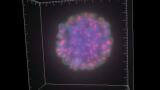
The DeltaVision OMX, on the other hand, gives the Department the ability to image at speeds of up to 100 frames/second with excellent spatial resolution.
ILM 51.5 July 2026

-(1).jpg)


-(1).jpg)
.jpg)