Laboratory products
Published over 16 years ago. See the latest and most current information on Laboratory products.
A top-of-the-range DeltaVision Core microscopy imaging system supplied by Preston-based Image Solutions Ltd or Imsol is at the heart of efforts being made by a team of researchers to unravel the mystery of how human cells turn cancerous.
The imaging system, which is designed to increase the ability to look at more probes and samples over longer periods of time than any competitive solution, is being used at the ManchesterInterdisciplinary Biocentre (MIB). This flagship research institute within The University of Manchester houses over 60 research groups which can draw on expertise in engineering, physical sciences, life sciences and medical sciences.
Imsol’s DeltaVision Core is central to the activities of Dr Dean Jackson, a cancer biologist within MIB’s life sciences section. He heads up a small group investigating the links between structure and function within
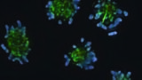
mammalian cells. In particular, the group is very interested in the regulation of cell division. Hence the need for an extremely powerful imaging solution that can handle both fixed and live cells.
According to Dr Jackson the two key advantages of the DeltaVision Core system are the speed and sensitivity that it brings to the group’s two main activities: live cell imaging and nuclear structure remodelling during differentiation.
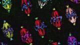
In the first, the imaging system is used to monitor the dynamic behaviour and stability of the structural subunits of the chromosomes known as DNA foci.
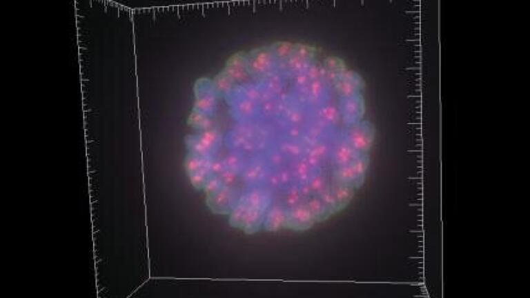
The second focus for the group involves the use of mouse and human cell models in an attempt to unravel the processes at work during cell differentiation. Here, the cells are dispersed in a complex 3D matrix in which they proliferate and form 3D balls. These balls eventually polarise, the central cells dying by apoptosis, to form hollow structures about 250 μm across known as acini. By labelling these cells, the scientists can use the DeltaVision Core for highly-detailed multi-channel imaging of the changes to 3D structure during the 14-21 day development and differentiation phase.
The DeltaVision Core has also established a reputation within the group of being easy to use.
ILM Guide 2026/27


.jpg)
.jpg)


.jpg)

.jpg)
.jpg)


-(1).jpg)


2.jpg)
