Microscopy & microtechniques
Published over 12 years ago. See the latest and most current information on Microscopy & microtechniques.
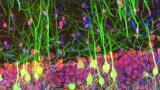
Multiphoton system FVMPE-R, the latest addition to the Olympus FluoView range of confocal laser scanning microscopes presents new possibilities in deep, in vivo imaging, creating high-resolution, three-dimensional images without damaging the sample.
The combination of laser scanning microscopy with fluorescence enables sharp image acquisition and measurement making it ideal for advanced imaging applications, from multicolour in vivo imaging to combined optogenetics (a technique using light to selectively activate ion channels in living specimens) and electrophysiology studies. Adaptable to fit individual custom-built systems, the FVMPE-RS can capture 438 fps at a image size of 512x32 px and at field number 18, tracking rapid processes taking place within the cell, tissue or organism in detail – from the transport of mitochondria through neurones to blood flow analysis or even Ca2+ signalling – all in real time.
For observing larger areas the system also captures full-frame, 512x512 px images at 30 fps made possible by a hybrid scanning unit, which builds on the capabilities of the high-speed resonant scanner. A galvanometric scanner is also available for maximising the signal to noise ratio during deep imaging. With the ability to seamlessly switch between the two,
Resolving the movement of the flowing bloodstream within a blood vessel is achieved at 438 fps under transmitted laser light observation.
An additional galvanometric scanner module, available for simultaneous 3D stimulation will assist with quantitative photomanipulation experiments and advanced optogenetics studies.This neuromodulation technique controls and monitors neuronal activity using light sensitive proteins channelrhodopsin and halorhodopsin, and with the FVMPE-RS, laser light stimulation of these optogenetic “switches” is achieved alongside simultaneous real-time-imaging of neuronal cell activity.
Also of importance in many biological processes are the interactions of cells within tissues or organs and the system can measure rapid fluctuations in groups of cells with a high signal-to-noise ratio using multipoint mapping. For example, this capability can generate information into how specific cell types contribute to the overall network function of an organ such as the brain.
Providing further insights into cell connectivity and how different cellular structures interact, the FVMPE-RS is optimised for tracking multiple molecular species simultaneously. Its four-axis alignment of multiple laser beams eliminates pixel shifts caused by excitation beam angle mismatches. This results in clear separation of multiple fluorophores enabling multiphoton excitation multicolour experiments. Reaching observation depths of up to 1.3 mm under in vivo conditions, depths of 8 mm can also be reached in non-living tissue treated with Scaleview tissue transparency reagent or by other methods.
ILM Guide 2026/27