Microscopy & microtechniques
Published over 14 years ago. See the latest and most current information on Microscopy & microtechniques.
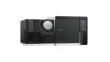
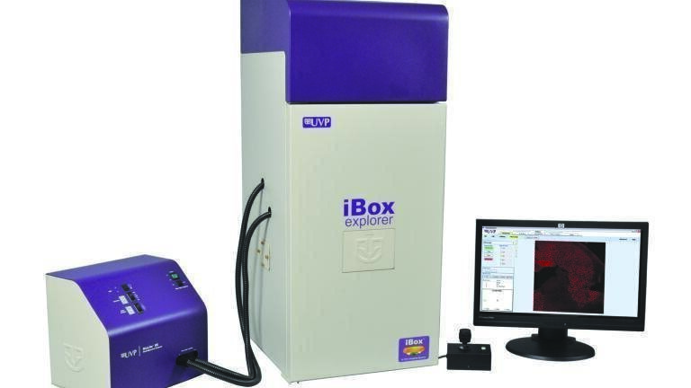
The iBox® ExplorerTM2 Imaging Microscope now features expanded capabilities for detection of fluorescent markers in small animals. With magnification from whole mouse to individual cells, the iBox Explorer2 provides breakthrough advances for cancer research studies. Researchers can now visualise tumour margins, micro metastasis and more with the wide range of magnifications from 0.17x to 16.5x. The upright optics provide an ultra-long working distance and high numerical aperture (NA) for detailed, exacting in vivo imaging research.
"Integrating the new optics further expands the instrument's current capabilities. Designed to transition from the macroscopic to the microscopic scale, the iBox Explorer2 permits deeper interrogation of a fluorescent signal in real time which otherwise would have necessitated cross-sectional studies," stated Antonio Sanchez, MD, UVP Life Scientist. "Scientists imaging whole animals for tumour tracking or biodistribution will be able to 'dive deeper' into the animal in order to investigate biological phenomena at the cellular level."
The iBox Explorer2 produces detailed images of tissues and cells with the leading-edge cooled scientific grade CCD camera. VisionWorks®LS software automates research with total system control and allows easy creation of templates for reproducible and consistent results.
The iBox Explorer2 utilises the BioLite™ Xe light source which provides bright illumination for multispectral fluorescent, visible and NIR excitation. The BioLite Xe houses a 150-watt xenon lamp that supplies brilliant excitation of fluorescent probes for a variety of applications. The BioLite Xe includes an eight-position emission filter wheel and convenient switching between experiments and multiplexing applications.
The iBox Explorer2 is the ideal system for in vivo imaging applications including: tumour shedding and angiogenesis; micro/macro metastases; tumour/host margins and interactions; tumour micro environment; primary tumour growth; hematogenous and intralymphatic trafficking; extravasation; biodistrubtion.
The iBox Explorer2 Imaging Microscope takes in vivo imaging to a new level with macro to micro subcutaneous to internal fluorescent imaging with one instrument.
ILM 51.6 Sept 2026


 Product directory


.jpg)