News
Published over 5 years ago. See the latest and most current information on News.
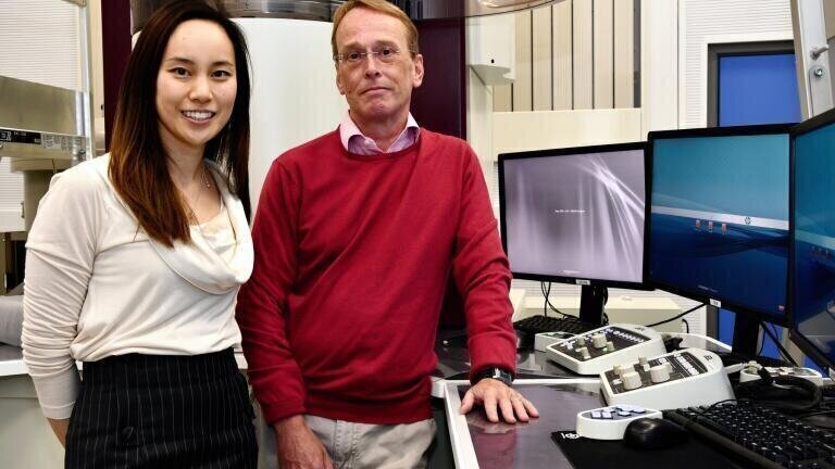
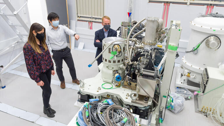
An electron microscope that can image biological samples at up to a million frames a second arrived at the Rosalind Franklin Institute on the Harwell Campus early in June. Shipped directly from Japan and the first of three instruments being developed jointly with JEOL Ltd., it will work with biological samples both cryogenically frozen and in liquids, which will enable imaging of molecules in motion.
By using speeds more commonly associated with imaging semi-conductors or catalysts (a thousand times faster than the current standard for biological samples) in combination with liquid cells the Correlated Imaging Team will use the Ruska microscope to ‘film’ proteins as they fold or to image drugs interacting with other molecules. For cryogenically frozen samples, the frames captured as the beam passes through the sample will enable the creation of 3D models of biological structures, such as viruses or proteins.
Correlated Imaging Deputy Director, Dr Judy Kim said: “We’ve already shown in initial experiments that we can use this technique to look at biological material. But the machine we were using wasn’t optimised for these kinds of samples. With the new machines, we’ll be able to refine the technique further. We’ll also be able to run very fast, to record data at close to a million frames a second, which will reduce the radiation damage visible on the sample.”
The machine, which will be located in a specialist EM suite minimising vibration, magnetic fields and acoustic noise and is also very carefully temperature controlled was amongst the first of equipment installations at the Rosalind Franklin building that will open fully later this year.
Correlated Imaging Director, Professor Angus Kirkland explained: “We’re looking at features that are smaller than the wavelength of normal visible light, so even tiny vibrations or changes in temperature can cause things to move. The machines have to be in a tightly controlled environment, so when we operate them, which we do remotely, there’s no risk of environmental interference.”
“The last five years have been very much about designing the instruments and building the platforms. But the next five years is about using those to do some really exciting science. We can’t wait to get started.”
Further information online
ILM 51.5 July 2026


.jpg)
.jpg)


-(1).jpg)


.jpg)