Scientific Imaging Competition Winners
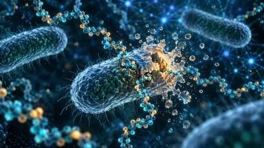
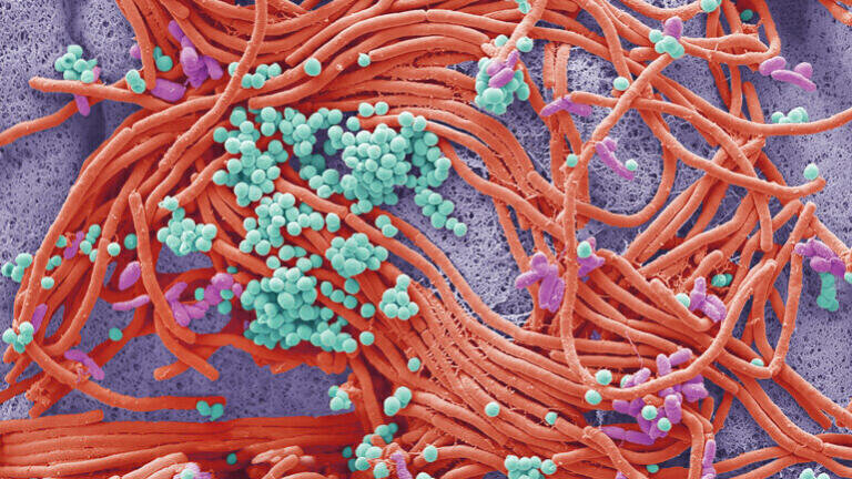
Electron Microscopy - Life Sciences 1st Prize Is your mobile bugged? SEM image of a Bacterial culture from a mobile phone. The average handset carries 18 times more potentially harmful germs than a flush handle in a men's toilet, tests have revealed. Steve Gschmeissner
Scanning Probe Microscopy
1st Prize
Light harvesting complex 2 (LH2)
Highest resolution image of single LH2 nonamer rings where defects in the ring are visible and single subunits can be discerned.
Sandip Kumar, University of Sheffield
Scanning Probe Microscopy 1st Prize Light harvesting complex 2 (LH2) Highest resolution image of single LH2 nonamer rings where defects in the ring are visible and single subunits can be discerned. Sandip Kumar, University of Sheffield
Light Microscopy - Physical Sciences 
1st Prize
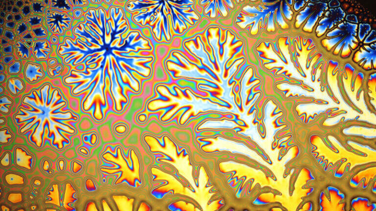
Pattern in a TiO2 sol-gel created by viscous fingers and light interference. Reflected light 200x magnification
In a sol-gel process, particles in a colloidal solution (sol) are converted to form a solid network (gel). Here, a spin-coated liquid sol film was covered by glass slide. Before solidifying air displaces the sol forming the viscous fingers pattern. The thin film interferes with visible light creating the colours.
Claire Trease, Kingston University
Light Microscopy - Physical Sciences 1st Prize Pattern in a TiO2 sol-gel created by viscous fingers and light interference. Reflected light 200x magnification In a sol-gel process, particles in a colloidal solution (sol) are converted to form a solid network (gel). Here, a spin-coated liquid sol film was covered by glass slide. Before solidifying air displaces the sol forming the viscous fingers pattern. The thin film interferes with visible light creating the colours. Claire Trease, Kingston University
Light Microscopy - Life Sciences
1st Prize
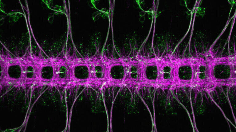
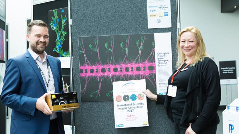
Drosophila melanogaster embryonic nervous system
Beautifully repeated segments of Drosophila melanogaster embryonic nervous system, stage 16/17, ventral view. Stained for Futsch (green, segmented sensory neurons and dispersed cell bodies in the ventral nerve cord) and Ank2-L (magenta, segmented ventral nerve cord and a subset of sensory neurons). Imaged with a Zeiss LSM780 confocal microscope.
Mohammad Mofatteh, MRC-Laboratory of Molecular Biology
Light Microscopy - Life Sciences 1st Prize Drosophila melanogaster embryonic nervous system Beautifully repeated segments of Drosophila melanogaster embryonic nervous system, stage 16/17, ventral view. Stained for Futsch (green, segmented sensory neurons and dispersed cell bodies in the ventral nerve cord) and Ank2-L (magenta, segmented ventral nerve cord and a subset of sensory neurons). Imaged with a Zeiss LSM780 confocal microscope. Mohammad Mofatteh, MRC-Laboratory of Molecular Biology
Electron Microscopy - Physical Sciences
1st Prize
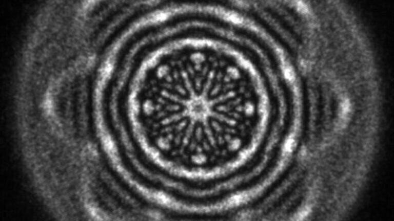
Shaped electron wavefunction
Zeroth order diffracted intensity of a structured electron beam generated using a focused-ion-beam-prepared holographic aperture comprising a superposition of two Laguerre-Gaussian states with quantum numbers L and p of (4,4) and (-4,2), respectively.        
Rafal Dunin-Borkowski, Forschungszentrum Jülich
Electron Microscopy - Physical Sciences 1st Prize Shaped electron wavefunction Zeroth order diffracted intensity of a structured electron beam generated using a focused-ion-beam-prepared holographic aperture comprising a superposition of two Laguerre-Gaussian states with quantum numbers L and p of (4,4) and (-4,2), respectively. Rafal Dunin-Borkowski, Forschungszentrum Jülich
People’s Choice Award
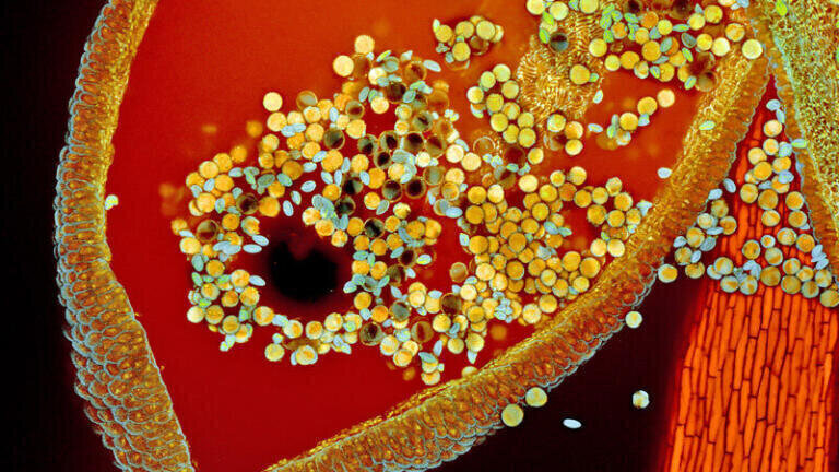
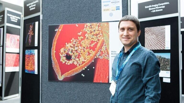
Hebe anther with pollen
Hebe anther with pollen scanned with confocal microscopy, using blue and red lasers. Auto-fluorescence was detected in four different channels, Zeiss 2012 Zen Black and Photoshop was used to enhance the colours. Water mounted sample, magnification 100x.            
Robert Markus & Zsuzsa Markus, University of Nottingham
People’s Choice Award Hebe anther with pollen Hebe anther with pollen scanned with confocal microscopy, using blue and red lasers. Auto-fluorescence was detected in four different channels, Zeiss 2012 Zen Black and Photoshop was used to enhance the colours. Water mounted sample, magnification 100x. Robert Markus & Zsuzsa Markus, University of Nottingham
Winning Short Video
Winning Short Video
Scientific Imaging Competition Winners

News

Scientific Imaging Competition Winners

16 Aug, 2017

Published over 8 years ago. See the latest and most current information on News.

This exciting international competition is a great opportunity for microscopists from across the world to showcase their skills. It provides a great outlet for those that can capture the breath-taking and engaging beauty of the microscopical world. The Society is always in awe of the quality of submissions across the categories.

With over 230 submissions, the 2017 RMS Scientific Imaging Competition was the largest yet. It's great to see so many people submitting their best work, but it made the judges lives very difficult. First, with the shortlisting process which gave us 50 images and videos to display at mmc2017, then with the final decisions on winners. So much so that a second prize in the video category had to be sought at the last minute!! Prize giving for the 2017 competition took place at mmc2017 in July.

We would like to thank the following companies for their generous sponsorship of the competition prizes:

Nikon Instruments UK

Carl Zeiss

Journal of Microscopy

Lambda

Agar Scientific

Bruker Nano Surfaces

Quekett Microscopical Club

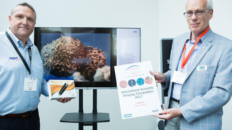
Many congratulations to all the winners including the short video category which was

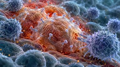
1st Prize -A nanoflight around a T-cell

Genetically engineered to fight lymphoblastic cancer. nanoflights are movies made in the scanning electron microscope with a very precise 8 axes piezostage and a multi-detector setup to get the coloured imaging. This flight had been the maybe smallest ever actually flown 360 in microscopic history. Diameter of T-cell is ca. 10-12 micron.

Stefan Diller

Latest News

ILM 51.5 July 2026

Explore our Digital Edition

Discover the latest news and research

Digital edition

Explore Our Other Sites

Envirotech Online
Instrumentation built for AMP8 and beyond
Explore more Arrow
Pollution Solutions Online
Keeping ballast water compliance on course
Explore more Arrow
Petro Online
Digitalisation advances at a large petrochemical complex in China
Explore more Arrow
Chromatography Today
Affordable liquid chromatography solvent delivery pump
Explore more Arrow