Research news
AI models have estimated the biological age of individual organs from tissue images and blood samples, with potential applications in disease monitoring and earlier diagnosis
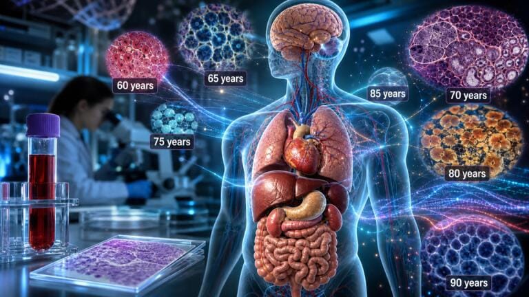
Artificial intelligence (AI) models known as ‘tissue clocks’ can estimate the biological age of human organs from microscopic tissue images, according to research led by scientists in Vienna.
Researchers analysed 25,712 images from 40 tissue types collected from 983 people. Their results indicated that organs did not age at a uniform rate and that tissue-specific signs of accelerated biological ageing could also be inferred from blood samples.
The study was led by the CeMM Research Center for Molecular Medicine of the Austrian Academy of Sciences and the Ludwig Boltzmann Institute for Network Medicine at the University of Vienna, Austria. Its authors said the findings could help scientists to investigate human ageing and might eventually support less invasive methods to assess organ health.
Chronological age records the time since a person’s birth, while biological age reflects the condition of the body and the aging effects of genetics, disease, lifestyle and environmental exposure. People of the same chronological age can therefore differ considerably in physiological health. Individual organs within one person might also follow distinct biological trajectories.
Previous biological clocks have often relied upon molecular measurements such as deoxyribonucleic acid methylation or gene expression. The Vienna-led team instead examined whether the physical architecture of tissue retained a record of age-related change.
The researchers used material from the Genotype-Tissue Expression Project. The collection included tissue from the brain, heart, lungs, kidneys, pancreas, adrenal glands, skin and intestine.
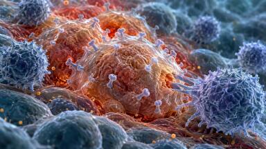
The team divided high-resolution tissue images into approximately 480 million smaller regions and applied computer vision models to identify subtle structural features. Age emerged as the strongest factor associated with tissue appearance across all 40 tissue types, although the models had not been explicitly instructed to search for it.
The researchers used these patterns to develop a separate tissue clock for each organ. Across the tissues studied, the clocks estimated age with a mean error of 4.9 years. The estimates were also associated with established features of ageing, including shorter telomeres, tissue abnormalities and a greater number of chronic diseases.
“Our tissues carry a remarkably detailed record of the ageing process,” said Dr. André Rendeiro, principal investigator at CeMM and corresponding author of the study.
“By combining histology images with AI, we can detect patterns of biological ageing that are invisible to the human eye,” he said.
The lungs, kidneys, pancreas and adrenal glands displayed signs of accelerated change between the ages of 20 and 40. Other tissues followed more complex courses and reached periods of faster change later in life. The uterus showed a marked shift at about the age of menopause.
Medical conditions were also associated with tissue-specific age estimates. Kidney failure was linked to accelerated biological-age signals in several tissues, while diabetes had a pronounced association with the pancreas.
“What stands out is how differently each organ ages and how that appears in tissue architecture,” said Dr. Ernesto Abila, a co-first author.
“Deep learning lets us read these spatial patterns and capture ageing as architectural remodelling, rather than only as molecular drift,” he added.
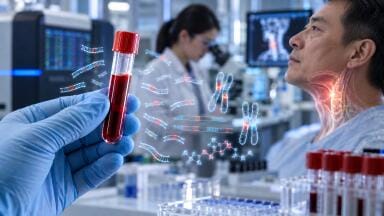
As tissue samples cannot always be collected safely, the team compared gene-expression profiles in blood with tissue-age gaps from the same individuals. It then developed predictors that could estimate the biological age of specific tissues from a blood sample alone.
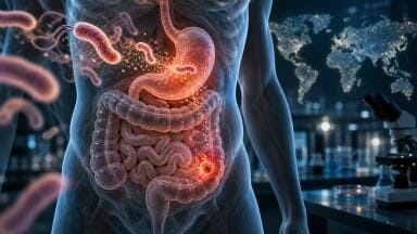
The blood-based models identified organ-age patterns associated with Alzheimer’s disease, Crohn’s disease, cystic fibrosis, vasculitis, diabetes and stroke. The strongest signal in Alzheimer’s disease appeared in the brain, while Crohn’s disease was associated with accelerated ageing across several parts of the gastrointestinal tract.
The method remains a research approach rather than a clinical diagnostic test. Further studies must establish whether tissue clocks can predict illness and produce reliable results across diverse populations.
If their clinical value is confirmed, blood-based tissue clocks could help doctors to monitor organ health without repeated biopsies, detect disease earlier and assess whether treatments have altered the biological condition of particular organs.
For further reading please visit: 10.1038/s41591-026-04566-5
ILM 51.6 Sept 2026


 Product directory









,-photo-Karin-Allander.jpg)




.jpg)