Fluorescence
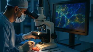
A research team at the University of Arizona has developed a light-based imaging technique that can distinguish healthy from cancerous pancreatic tissue within minutes using a combination of multiphoton microscopy with artificial intelligence and could allow surgeons to make real-time decisions during operation
Pancreatic neuroendocrine neoplasms (PNENs) are a rare cancer that affects the hormone-producing cells of the pancreas. Although uncommon, their incidence has been increasing for several decades. Treatments include chemotherapy and targeted therapies although surgery remains the only current curative option. Surgical outcomes, however, often depend on pathology results that can take many hours or even days to confirm, which delays clinical decisions and raises the risk of incomplete tumour removal.
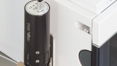
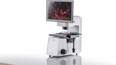
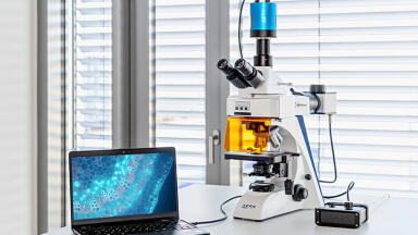
Now, a research team at the University of Arizona, in Tucson, has reported the development of a novel imaging technique that could enable surgeons to identify cancerous tissue more quickly and accurately. The method – called multiphoton microscopy (MPM) – uses a light-based imaging approach to visualise naturally fluorescent molecules within tissue. Unlike conventional microscopy, MPM causes less structural damage and yields clearer images, which makes it a promising candidate for real-time use during surgery.
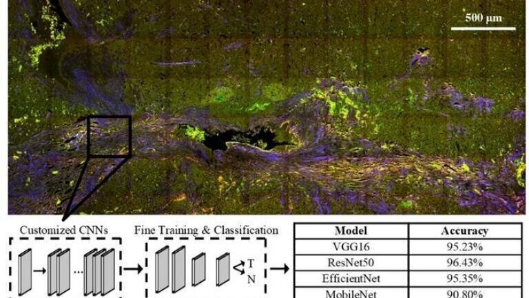
The researchers applied MPM to pancreatic tissue samples to visualise naturally occurring fluorescent markers such as collagen, nicotinamide adenine dinucleotide, flavin adenine dinucleotide, lipofuscins and porphyrins. These compounds serve as intrinsic identifiers that allow pathologists to distinguish between healthy and malignant tissue. To interpret the images, the researchers trained one machine learning (ML) algorithm and four convolutional neural networks (CNNs) to classify the samples by tissue type.
The ML algorithm achieved an accuracy of 80.6 percent in identifying cancerous tissue, while the CNNs performed even better, with accuracies from 90.8 percent to 96.4 percent. These strong results are particularly notable because the samples originated from several independent biorepositories which suggests that the method is robust across diverse data sources.
Although the CNNs achieved higher performance, the ML model offered greater transparency. By examining the features that most influenced the algorithm’s decisions, the researchers identified collagen content and image properties such as contrast and correlation as the principal discriminators between healthy and cancerous tissue. These insights could help refine subsequent models and deepen understanding of the structural changes characteristic of PNENs.
The study also demonstrated that MPM imaging is faster than traditional histological assessment, although the team believes that further refinement could improve speed and clinical usability. The researchers next intend to evaluate the technique on fresh tissue samples during surgery and to determine whether it can accurately assess tumour grade and subtype, information that would guide treatment planning with greater precision.
This research indicates that intraoperative cancer diagnosis and surgical planning may soon occur in near real time, potentially reducing the need for secondary procedures and improving outcomes for patients with pancreatic cancer.
For further reading please visit: 10.1117/1.BIOS.2.4.045001
ILM 51.5 July 2026


-(1).jpg)
.jpg)


-(3).jpg)