

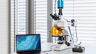
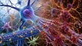
Fluorescence
Binding-dependent fluorescent nanobody probes have delivered high-specificity, low-noise imaging across the visible spectrum, with applications from neuroscience to developmental biology
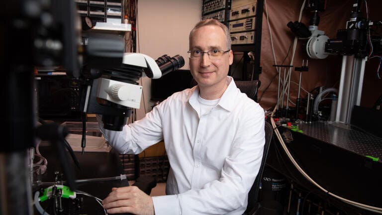
Fluorescent proteins have transformed modern biology by enabling scientists to label and track individual molecules within living cells, tissues and whole organisms. These tools have allowed researchers to observe viral infection in real time, to examine intracellular degradation pathways and to follow signalling cascades that drive tumour progression. Now, investigators at the Salk Institute for Biological Studies, La Jolla, California, USA, in collaboration with the Albert Einstein College of Medicine, Bronx, NYC, USA, have reported a refined imaging platform designed to improve signal clarity and specificity in complex biological systems.
The technology – visible-spectrum antigen-stabilisable fluorescent nanobodies, or VIS-Fbs – has been validated across multiple mammalian cell types and has demonstrated broad utility in live-cell and in vivo imaging. The approach builds on nanobody-based probes which are derived from small antibody fragments that can be engineered to bind specific intracellular targets. When fused to fluorescent proteins, these probes have enabled researchers to visualise protein localisation and dynamics within living systems. However, conventional constructs have continued to emit fluorescence even in the absence of target binding, which has introduced background signal and reduced image fidelity.
The present work has addressed this limitation through a binding-dependent fluorescence mechanism. VIS-Fbs remain unstable and non-fluorescent unless bound to their intended antigen. Upon binding, the probe stabilises and emits a fluorescent signal. This conditional activation has reduced background fluorescence by approximately one hundredfold, allowing substantially clearer visualisation of molecular events at high spatial resolution. The effect has been particularly marked in densely populated or optically challenging environments, where conventional probes often struggle to distinguish signal from noise.
“This work establishes a versatile platform for imaging proteins with high specificity and minimal background,” said the research team leaders Dr. Axel Nimmerjahn, professor and Dr. Françoise Gilot-Salk Chair at the Salk Institute.
“It opens new opportunities to study how molecular and cellular processes unfold in real time across diverse biological systems,” they added.
The researchers have also expanded the spectral range of the probes. Multiple VIS-Fb variants have been engineered to fluoresce across nearly the full visible spectrum, from blue wavelengths through to far red emission. This spectral diversity allows simultaneous imaging of multiple targets within the same cell or tissue, which is essential for dissecting complex signalling networks and multi-component biological processes.
In addition, selected variants have incorporated photoswitchable properties, which enable researchers to toggle fluorescence on and off with light. This feature supports longitudinal tracking of protein behaviour with both high temporal resolution and precise spatial control.
A further strength of the system lies in its modular architecture. The design framework allows rapid adaptation of the probes to bind different molecular targets or to incorporate functional reporters. This flexibility has positioned VIS-Fbs as a generalisable platform rather than a single-purpose tool, with potential applications spanning cell biology, neuroscience and translational research.
Experimental validation has extended beyond cultured cells to include whole-organism models. In murine systems, VIS-Fb probes have enabled selective labelling and ratiometric imaging of calcium dynamics in both neurons and astrocytes during behavioural activity. These measurements have provided insight into cell-type-specific signalling within intact neural circuits. In zebrafish, the probes have allowed real-time observation of developmental processes and responses to pharmacological perturbation, offering a dynamic view of signalling pathway modulation during early life stages.
“Our results show that this imaging platform offers a much clearer and more precise view of how proteins behave inside living systems,” said Dr. Vladislav Verkhusha, professor and co-director of the Gruss Lipper Biophotonics Center at the Albert Einstein College of Medicine.
“It opens the door to study complex biological processes, such as cell signalling, development and disease progression, in novel ways,” he said.
The development of VIS-Fbs reflects a broader trend within bioimaging to increase both sensitivity and specificity while maintaining compatibility with living systems. By minimising background fluorescence and expanding multiplexing capability, the platform has the potential to support more accurate interpretation of intracellular dynamics, particularly in heterogeneous tissues such as the brain. As imaging technologies continue to evolve, tools that reduce artefact and enhance signal fidelity are likely to play a central role in efforts to map biological function at molecular resolution.
For further reading please visit: 10.1038/s41592-026-03056-3
Lab Asia 33.4 - August 2026


-(1).jpg)


-(3).jpg)


-(1).jpg)
