Laboratory products
Published over 14 years ago. See the latest and most current information on Laboratory products.
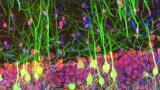
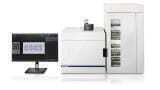
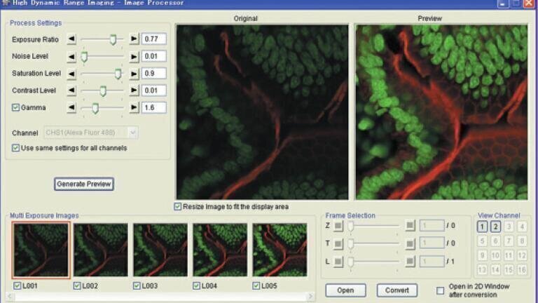
Olympus has introduced FV10-ASW 3.0, the latest version of its software for the FluoView FV1000 range of confocal laser scanning microscopes (cLSM) and Fluoview FV1000MPE (multiphoton excitation) systems. By incorporating high dynamic range imaging (HDRI), a greater dynamic range of fluorescence between the lightest and darkest areas of a sample is obtained. Furthermore, signal to noise ratios are minimised, enabling clear, highly resolved images to be easily captured. Additional functionality, such as partial stitching with multi area time lapse imaging, and channel unmixing for the FluoView FV1000MPE systems, provides advanced flexibility for obtaining the best possible LSM images, every time. The new software is also fully compatible with Microsoft Windows 7. As an advanced image composition technique, HDRI provides users with a powerful tool to achieve high-quality images with outstanding contrast and no saturation. By effectively merging multiple images captured under different exposure settings, the final image provides outstanding clarity, with the ability to visualise even the more challenging cellular components. With advanced multi area time-lapse functionality, version 3.0 of the FV10-ASW software allows users to select specific areas of the whole sample, which can be chosen and stitched together with ease. This time saving function is key in allowing the quick and simple analysis of specific regions of interest.
With specific advances for the FV1000MPE systems, the non-descanned detectors, which enable users to cover every multiphoton excitation requirement and acquire maximum fluorescence output, can now also be used for channel un-mixing. As a result, users can perform spectral un-mixing of multi-colour samples, even those with significant spectral emission cross over. A laser limiter also sets upper intensity boundaries to avoid over stimulation of sensitive samples, thus ensuring that cellular integrity is maintained. The Olympus FluoView FV1000 microscope systems incorporate a unique SIM scanner concept with two independent, fully synchronised laser scanners in a single, compact design. This enables simultaneous, high-resolution confocal observation. The advanced optical detection system of the FV1000 provides superior linear spectral distribution throughout the wavelength range of 400 to 800nm. Based on the FluoView FV1000 cLSM, the FV1000MPE system for deep in vivo imaging of live and thick slice samples is available in three different models. Each incorporates Olympus’s market leading components such as the UIS2 optics. The systems are fully configured to ensure the best excitation efficiency, allowing accurate imaging hundreds of microns deep into a specimen. Researchers can therefore achieve in vivo optical sectioning, without causing any significant photobleaching or phototoxicity.
ILM 51.5 July 2026


-(1).jpg)
.jpg)

.jpg)
.jpg)