Research news
Large-scale imaging study reveals that skeletal muscle quality and fat distribution outperform BMI in predicting diabetes, cardiovascular events and mortality
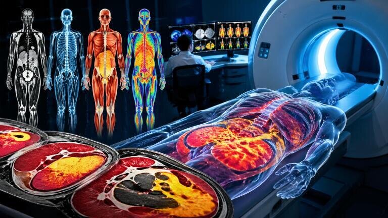
A research team of radiologists has developed a detailed reference map of human body composition – with the use of artificial intelligence – to analyse whole-body MRI scans from more than 66,000 participants. The work has shown that both the quantity and quality of skeletal muscle serve as strong predictors of diabetes, major cardiovascular events and mortality, with implications that extend beyond traditional reliance on indicators such as body mass index (BMI).
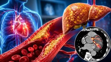
Clinicians have long used BMI to estimate cardiometabolic risk, which describes the interaction between cardiovascular health and metabolic processes such as energy use and nutrient handling. However, BMI remains a crude proxy, since it considers only height and weight and does not capture the distribution of adipose tissue or the composition of muscle.
“Many risk scores and treatment decisions still rely on BMI or waist circumference because they are simple to obtain,” said Dr. Jakob Weiss, senior author and radiologist at the department of diagnostic and interventional radiology at the University Medical Center, Freiburg, Germany.
“But BMI does not reliably reflect a person’s actual body composition, he said.”
The absence of robust reference standards for body composition across the lifespan has limited clinical interpretation. Ageing, sex and height all exert measurable effects on fat deposition and muscle structure, yet comparative benchmarks for asymptomatic populations have remained underdeveloped.
“There is growing evidence that body composition measures are independent risk factors for cardiometabolic and oncological diseases and mortality,” said Dr. Matthias Jung, first author from the same institution.
“However, these measures are influenced by height and sex and change substantially with age,” he added.
The retrospective analysis drew on 66,608 individuals with a mean age of 57.7 years, including 34,443 male participants and a mean BMI of 26.2. All participants had undergone whole-body magnetic resonance imaging as part of large-scale population studies, including the UK Biobank and the German National Cohort, between April 2014 and May 2022.
Researchers applied an open-source, fully automated deep learning framework to extract quantitative metrics from imaging data. These metrics included subcutaneous adipose tissue, visceral adipose tissue, skeletal muscle volume, skeletal muscle fat fraction and intramuscular adipose tissue. Each measurement was normalised for age, sex and height and expressed as a ‘z-score’, which indicates the degree of deviation from population-adjusted norms.
Subsequent statistical analysis assessed how these z-score categories predicted clinical outcomes. Individuals with high visceral fat showed a 2.26-fold increased risk of future diabetes. Elevated intramuscular fat correlated with a 1.54-fold increased risk of major adverse cardiovascular events, while low skeletal muscle mass associated with a 1.44-fold increase in all-cause mortality, independent of established cardiometabolic risk factors.
“It’s not only how much muscle you have but, also, it’s the quality of that muscle. Knowing the volume of intramuscular fat gives us a window into muscle quality that other methods like BMI, bioelectrical impedance analysis, or dual-energy X-ray absorptiometry cannot easily provide,” Jung said.
Beyond risk prediction, the team has constructed age-, sex- and height-adjusted reference curves for each key metric. These curves establish a comparative framework that allows clinicians to determine whether an individual’s body composition deviates significantly from expected norms.
“Adjusting for confounding factors is critical to improve screening accuracy and tailor treatment decisions,” Weiss said.
“This tool has the potential to identify whether an individual’s body composition puts them at greater risk for metabolic disease compared to their age-matched peers,” he added.
To facilitate adoption, the researchers have released an open-source, web-based calculator that generates normalised z-scores for body composition. The platform has aimed to support standardisation across studies and clinical settings, with the goal to improve comparability and external validity.
“This tool can allow clinicians to use routine imaging opportunistically,” Weiss said.
“A dedicated whole-body MRI is not necessarily required. If a routine CT or MRI body scan already exists, the information can be extracted for benchmarking against the reference values,” he said.
The approach may extend beyond cardiometabolic disease. The authors suggested that refined body composition metrics could enhance risk stratification in oncology and help clinicians distinguish between beneficial fat loss and detrimental muscle loss in patients who receive glucagon-like peptide-1 receptor agonists for weight management.
“We’re already imaging patients every day. On every scan of the abdomen or chest, the information is there, we just don’t routinely measure or report it. AI now allows us to tap into this hidden layer of data in a quantitative [and] reproducible way,” Dr Weiss said.
The team has indicated that future work will focus on validation of these reference curves in clinical populations, with particular emphasis on prediction of treatment toxicity, survival outcomes and disease recurrence in cancer cohorts. Researchers have also planned to derive disease-specific reference standards tailored to distinct patient groups.
For further reading please search for:
Body Composition in the General Population: Whole-body MRI-derived Reference Curves from Over 66,000 Individuals
ILM Guide 2026/27


2.jpg)
