Research news
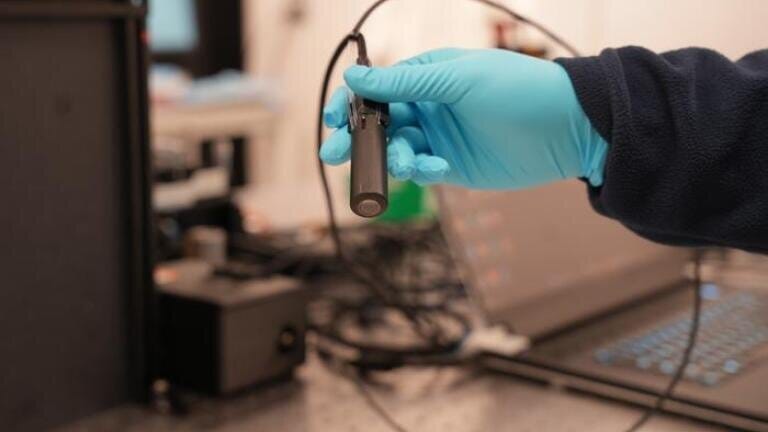
Rice University and MD Anderson researchers have developed PrecisionView, a handheld imaging device that combines artificial intelligence-designed optics with real-time tissue imaging to help clinicians detect signs of cancer without immediate reliance on biopsy
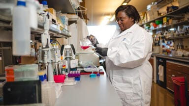
Researchers at Rice University and The University of Texas MD Anderson Cancer Center both in Houston, Texas, USA, have developed a compact, artificial intelligence (AI)-powered imaging device intended to help clinicians detect cancer earlier and more accurately at the point of care.
The technology – called PrecisionView – is a handheld endomicroscope which combines advanced optical design with a subset of machine learning called ‘deep learning’ to allow clinicians to view cellular structures and underlying blood vessels across large tissue areas in real time. The researchers said the device could reduce dependence on invasive biopsies by helping clinicians to examine broader regions of tissue and identify areas most likely to require further investigation.
“Early detection is one of the most critical factors in improving cancer outcomes but today’s tools often force clinicians to choose between detail and coverage,” said Dr. Rebecca Richards-Kortum, the ‘Malcolm Gillis’ university professor at Rice and co-director of the Rice360 Institute for Global Health Technologies and corresponding author for the study.
“With PrecisionView, we no longer have to make that trade-off – we can see both clearly and in real time,” she said.
Epithelial cancers, including cancers of the cervix and oral cavity, account for an estimated five per cent of all cancer cases worldwide. Many are still diagnosed at a late stage, in part because diagnostic pathways still rely heavily on biopsy. While biopsy remains essential to gain a definitive diagnosis, by definition it can sample only a limited region of tissue and may miss clinically important changes elsewhere in a lesion.
Traditional in vivo microscopy can offer a less invasive alternative but it is constrained by a restricted field of view, limited depth of field and difficulty with uneven tissue surfaces. These limitations can make it harder to assess large or irregular lesions and to decide where biopsy is most appropriate.
PrecisionView addresses these limitations through a deep learning-optimised optical system and real-time image reconstruction. About the size of a large pen, the device uses a custom-designed phase mask and an AI reconstruction algorithm to expand its imaging capability. According to the researchers, it achieved a field of view about five times larger and a depth of field about eight times greater than conventional systems while retaining cellular-level resolution.
“Traditionally, machine learning and AI tools are used to enhance images in terms of resolution or contrast, after the images have been acquired by conventional imaging systems,” said Dr. Ashok Veeraraghavan, chair of electrical and computer engineering at Rice and co-author of the study.
“In stark contrast, this work [uses an] AI approach to redesign the optics of a microscope. The AI-designed optics not only improves resolution and contrast, but more importantly breaks the conventional trade-off between depth of field and resolution – creating a handheld microscope platform that still achieves cellular resolution, while providing an eight-times increase in depth of field.”
He added that the improvement in depth of field was critical for real-world use because it made it practical for clinicians and technicians to hold the device by hand and obtain high-resolution images without focal blur compromising image quality.
The device allows clinicians to view two important features associated with cancer in a single image:
These structural and vascular changes can help clinicians to distinguish healthy tissue from precancerous or cancerous lesions.
“Being able to capture both nuclear and vascular features in a single, continuous image is a major step forward, because these are the signals clinicians rely on to distinguish healthy tissue from precancerous or cancerous lesions,” said Dr. Huayu Hou, a postdoctoral associate in Richards-Kortum’s Optical Spectroscopy and Imaging Laboratory and one of the paper’s authors.
PrecisionView can generate detailed tissue maps across areas that span several square centimetres and display results in real time at up to 15 frames per second. The researchers validated the device through experiments that included imaging of healthy volunteers and human tissue samples with precancerous lesions. In one study, the device was used to scan the oral cavity of volunteers and produced high-resolution maps of tissue structure and blood vessels across areas larger than one square centimetre. In another, it identified precancerous changes in cervical tissue and distinguished abnormal regions from surrounding healthy tissue.
“Instead of sampling a small piece of tissue and sending it to a lab, this technology allows us to assess a much larger area instantly,” said Dr. Jimin Wu, a postdoctoral associate in electrical and computer engineering and one of the authors of the study.
“That could significantly reduce missed diagnoses and unnecessary procedures,” she added.
The researchers said accessibility had been central to the device’s design. PrecisionView has been built with relatively simple components and costs around US$3,000, which could make it suitable for clinics and lower-resource settings where conventional pathology infrastructure is limited. Low-cost health care technologies for global use are a major focus of Rice360.
“PrecisionView has the potential to bring high-quality diagnostic capability directly to the point of care – helping clinicians make more timely decisions which will improve access to life-saving early detection,” said Dr. Kathleen Schmeler, associate vice president of global oncology in the Department of Cancer Network, Division of Surgery at MD Anderson and one of the authors of the study.
“The impact will be particularly significant in medically underserved areas where access to pathology services may be limited or delayed, leading to missed or late diagnoses,” she said.
The team said the technology could support clinical applications that include biopsy guidance, surgical decision-making and earlier cancer detection during routine screening. However, the researchers emphasised that larger clinical studies would still be needed to validate diagnostic accuracy before the device could be used widely in routine care.
“PrecisionView represents a future direction for medical imaging, one where AI and optical design work together to improve outcomes,” Dr. Richards-Kortum said.
“By designing hardware and algorithms together, we can unlock capabilities that simply weren’t possible before,” he concluded.
For further reading please visit: 10.1073/pnas.2602705123
ILM Guide 2026/27


2.jpg)
