Research news
A formulation of methylene blue delivered via dissolvable microneedles has improved lymphatic imaging clarity, targeting and functional assessment, with the potential to advance treatment of lymphatic disorders
A research team from China has reported a minimally-invasive imaging strategy that may address long-standing limitations in the assessment of the lymphatic system, the network central to fluid homeostasis, immune surveillance and disease progression. By combining dissolvable microneedles with a nanoparticle formulation of methylene blue, the team has achieved enhanced visualisation of lymphatic vessels and nodes alongside the ability to observe functional dynamics in vivo.
The lymphatic system plays a pivotal role across a spectrum of pathological conditions, from lymphoedema and chronic inflammation to tumour metastasis, venous insufficiency and impaired wound repair. Accurate evaluation of both its structure and function has therefore become increasingly important in clinical and translational settings. However, existing imaging modalities such as lymphoscintigraphy, magnetic resonance lymphangiography, indocyanine green fluorescence imaging and conventional methylene blue imaging have remained constrained by trade-offs that include invasiveness, limited spatial resolution, poor targeting specificity, radiation exposure and high operational cost.
Although methylene blue has long served as a clinically approved near-infrared dye with a favourable safety profile, its utility in lymphatic imaging has been restricted by physicochemical limitations. The molecule tends to aggregate in aqueous environments and – owing to its small particle size – shows limited lymphatic selectivity. At the same time, standard intradermal injection techniques required to deliver such tracers can cause discomfort and rely heavily on operator skill which can introduce diverse variability in clinical practice.
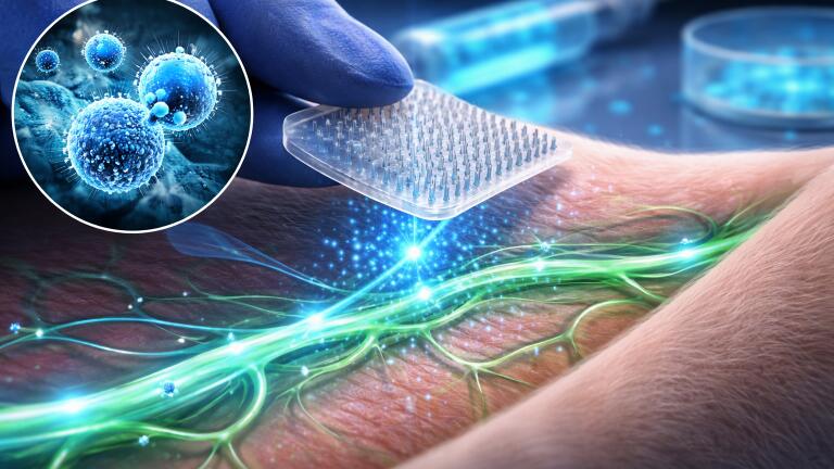
Against this background, investigators from Shanghai Ninth People’s Hospital, Shanghai Jiao Tong University School of Medicine, together with collaborators from Shanghai Jiao Tong University and Shanghai Children’s Medical Center, China, have developed a nanoparticle-based formulation designed to overcome these constraints. The work has demonstrated that methylene blue encapsulated within an MPEG-PCL nanocarrier and delivered through dissolvable microneedles has enabled near-infrared imaging of lymphatic structures with improved clarity, targeting and functional insight when compared with conventional approaches.
To construct the platform, the researchers encapsulated methylene blue within a methoxy polyethylene glycol–polycaprolactone carrier using a double-emulsion method. This approach increased the effective particle diameter from less than 10 nanometres in the free dye to approximately 99 nanometres, a size range that is more favourable for lymphatic uptake. The nanoparticle formulation also exhibited a modest negative surface charge, enhanced fluorescence intensity and improved resistance to quenching and degradation in aqueous conditions, all of which contribute to more stable imaging performance.
In vitro experiments have demonstrated that the material exhibited low cytotoxicity and caused haemolysis of less than five per cent, while cellular assays confirmed uptake and transendothelial transport across lymphatic endothelial cells. These findings have supported its candidacy as a targeted lymphatic imaging agent with acceptable biocompatibility.
The delivery mechanism represents a further key component of the system. The team incorporated the nanoparticle tracer into an array of dissolvable microneedles arranged in a 15×15 configuration. Mechanical testing has shown that these microneedles possessed sufficient strength to penetrate the stratum corneum – the outermost layer of the epidermis – while in vitro release studies indicated that approximately 80 per cent of the payload was delivered after insertion. By confining delivery to the superficial dermis, the system has avoided activation of deeper nociceptors, which has enabled near-painless administration without reliance on conventional hypodermic injection.
In animal models, the combined platform has outperformed established comparators, including indocyanine green and unmodified methylene blue. Imaging studies in rats have demonstrated that signal intensity reached at least threefold higher levels than those achieved with conventional tracers at equivalent concentrations, resulting in markedly improved delineation of lymphatic vessels. The approach has also reduced extravasation at the injection site, which has allowed clearer identification of dominant lymphatic pathways and nodal structures.
Perhaps most notably, the method has enabled visualisation of rhythmic segmental contractions along lymphatic vessels. These contractile events, which underpin lymph propulsion, have historically proven difficult to capture using non-invasive imaging. Their detection here has suggested that the technique can extend beyond static anatomical mapping to provide insight into lymphatic function in real time.
“This study turns a familiar clinical dye into a smarter lymphatic probe,” the researchers stated. By integrating nanoscale engineering with a minimally invasive delivery system, the work has addressed multiple technical barriers simultaneously, including weak lymphatic targeting, signal instability, procedural discomfort and limited capacity to assess functional dynamics.
A portable, nonradioactive and minimally invasive imaging platform could support earlier detection of lymphatic dysfunction, improve longitudinal monitoring of lymphoedema and refine evaluation of diseases in which lymphatic transport is compromised. The authors have also reported that the tracer demonstrated favourable biosafety profiles in vivo, with no significant pathological changes observed in major organs and routine blood parameters remaining within normal ranges. This raises the possibility of repeated or longer-term use as the technology advances towards clinical translation.
With further optimisation and validation in human studies, the platform may help to bring lymphatic imaging closer to routine bedside application.
For further reading please visit: 10.1093/burnst/tkaf067
ILM Guide 2026/27


2.jpg)
