Research news
Researchers at Johns Hopkins Medicine have demonstrated that modification of a key retinal barrier has improved survival, migration and functional maturation of transplanted nerve cells, with implications for future vision restoration therapies
A research team at Johns Hopkins Medicine, Baltimore, Maryland, USA has reported that disruption of a long-suspected structural barrier within the eye has enabled transplanted nerve cells to survive, migrate and begin to integrate into retinal tissue, a finding that may help to restore vision in patients with optic nerve damage.
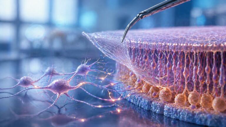
The study – funded by the National Institutes of Health – has examined how alteration of the internal limiting membrane affects the fate of transplanted retinal ganglion cells. This thin, specialised layer separates the retina from the vitreous humour and was thought to impede attempts to replace neurons lost in optic neuropathies.
Optic neuropathy arises when retinal ganglion cells fail, commonly as a result of conditions such as glaucoma, optic neuritis or ischaemic optic neuropathy. These cells play a central role in vision, as they transmit electrical signals from the retina to the brain. They do not regenerate naturally and so cell replacement strategies have become a major focus within regenerative ophthalmology.
The research team used a combination of animal models, laboratory-grown human cells and donated ocular tissue to investigate whether the internal limiting membrane constitutes a physical barrier to successful transplantation. Previous hypotheses have proposed such a role, but direct in vivo evidence has been limited.
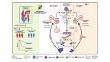
To test this, the investigators introduced human retinal ganglion cells into three groups of immunosuppressed mouse models. One group carried a genetic mutation that produced an incomplete, discontinuous internal limiting membrane. A second group received an enzyme treatment designed to partially digest the membrane without damage to surrounding tissue. A third group acted as a control and received an inert solution.
After two weeks, survival of transplanted cells reached 95 per cent in eyes with the structural defect, compared with 80 per cent in enzyme-treated eyes and 75 per cent in control animals. More importantly, the researchers observed that disruption of the membrane allowed a substantially greater proportion of transplanted cells to enter the retinal ganglion cell layer, where they need to be to contribute to visual function.
Three-dimensional imaging provided further evidence of functional integration. Between two per cent plus or minus 0.6 per cent and 7.1 per cent plus or minus 1.6 per cent of surviving cells in enzyme-treated and mutant eyes, respectively, developed dendritic processes. These structures are essential for neuronal communication and signal processing. By contrast, only 0.01 per cent plus or minus 0.01 per cent of cells in control eyes showed comparable maturation, which has reinforced the view that the intact membrane limits both migration and differentiation.
Parallel experiments conducted in larger animal eyes and in donated human retinal tissue have reproduced these findings, which the authors have said provides compelling evidence that the internal limiting membrane represents a critical structural barrier to neuronal replacement strategies.
The team has also established a reproducible surgical approach to transplant retinal ganglion cells in conjunction with controlled disruption of the membrane. This procedural framework may support future clinical trials, where safe and effective integration of transplanted neurons remains a central challenge.
“Even when the retinal ganglion cells survive, they remain on the retina’s surface and do not migrate into the tissue or form the connections with other nerve cells necessary to detect light,” said Dr. Thomas Vincent Johnson III, the ‘Shelley and Allan Holt Rising Professor of Ophthalmology’ at the Wilmer Eye Institute in Baltimore.
The findings have advanced the field by shifting focus towards the physical microenvironment of the retina rather than solely the intrinsic properties of transplanted cells. However, the investigators have emphasised that substantial translational work remains before such strategies reach clinical practice.
“We know our methods are effective but we do not know if completely removing the internal limiting membrane helps or harms the retinal ganglion cells in the long run,” Johnson said.
“It will likely take several years before our findings could become available as an experimental therapy, but the methods we developed will guide the field moving forward,” he added.
Taken together, the study provides a clearer mechanistic understanding of why retinal ganglion cell transplantation has historically failed and it has identified a tangible target for intervention. If confirmed in human trials, controlled modification of the internal limiting membrane may become a foundational step in efforts to restore vision in patients affected by optic nerve degeneration.
For further reading please visit: 10.1126/scitranslmed.adr1062
ILM Guide 2026/27


2.jpg)
