Research news
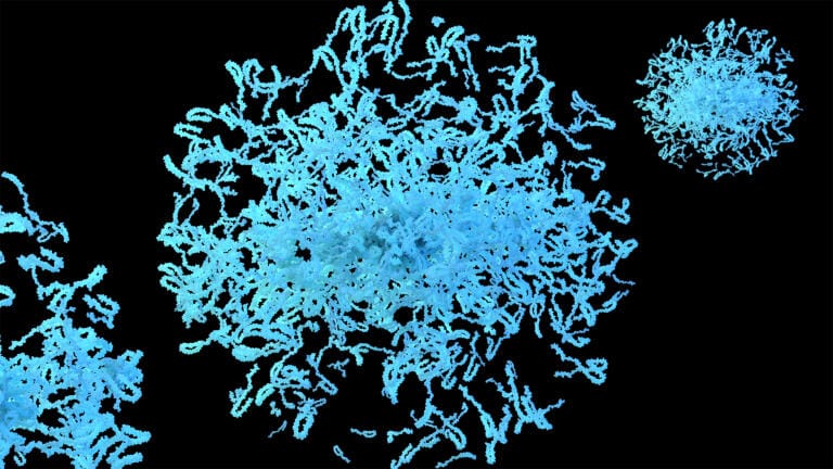
Scientists have demonstrated, for the first time, the ability to directly visualise pathological alpha-synuclein aggregates in the living human brain using a newly developed PET tracer. The advance could improve the diagnosis of neurodegenerative disorders and accelerate the development of targeted therapies.
Developed by MODAG in collaboration with the University Hospital Tübingen and the Max Planck Institute for Multidisciplinary Sciences, the tracer – [11C]MODAG-005 – selectively binds to abnormal alpha-synuclein deposits associated with Parkinson’s disease, multiple system atrophy and dementia with Lewy bodies. The findings [1] have been published in Science Translational Medicine.
Alpha-synuclein aggregates are believed to accumulate years before clinical symptoms appear, yet until now they have remained largely inaccessible to direct imaging in living patients. The new tracer enables these disease-associated protein deposits to be detected using positron emission tomography (PET), offering a potential route to earlier and more accurate diagnosis.
Alpha-synuclein is widely regarded as a key driver of several neurodegenerative disorders, but tracking its accumulation in living patients remains a major challenge. Current diagnoses often rely on clinical symptoms, which can overlap between diseases and may only emerge after significant neurological damage has occurred. Direct visualisation of the protein could therefore provide a powerful tool for both research and patient care.
“With this new approach, we can observe pathological processes directly in the brain well before clinical symptoms become apparent,” said Professor Kristina Herfert of the Werner Siemens Imaging Center at University Hospital Tübingen. “This opens new possibilities for earlier and more accurate diagnosis.”
The researchers validated the tracer through laboratory studies, animal models and initial patient imaging. Early results showed high binding specificity and produced detailed images of disease-related pathology. The team also found evidence that different neurodegenerative diseases may generate distinct patterns of alpha-synuclein accumulation, raising the possibility of biology-based differential diagnosis.
Beyond diagnosis, the technology could become a valuable tool for drug development, allowing researchers to determine whether experimental therapies engage their intended target in the brain. This may help improve the design and evaluation of clinical trials for disease-modifying treatments.
MODAG is also developing a fluorine-18-labelled version of the tracer, designed to expand availability beyond specialist imaging centres and support future use in routine clinical practice.
More information online
ILM Guide 2026/27


2.jpg)
