Mass Spectrometry & Spectroscopy
Protein Characterisation Techniques: How to Characterise a Protein
Jul 20 2021
Made up of multiple chains of amino acids, proteins are complex molecules that play a critical role in cellular growth, maintenance, and repair. While all proteins share some core similarities, variables like size, physiochemical properties, amino acid sequence and molecular structure can vary enormously. This is where protein characterisation techniques step up. Used to detect, isolate and map the unique amino acids that make up a protein chain, techniques like Mass Spectrometry and Dynamic Light Scattering allow scientists to profile proteins in enormous detail.
Below, we explore some of the most useful protein characterisation techniques used in laboratories around the world. We also spotlight some key applications for protein characterisation and explore why the field is critical to modern science.
Proteins: 101
Before we dive in, let’s take a moment to define proteins. Large and complex, proteins are three-dimensional molecular structures folded into unique shapes. Each protein is made up of smaller units called amino acids, which are bound together with peptides to form a long chain. While there are only 20 different types of amino acids found in proteins, each chain can feature hundreds and sometimes thousands of individual amino acids. Sequence is important, with the order of the amino acids determining the three-dimensional structure and unique function of a protein. To summarise, the unique characteristics of a protein are determined by the amino acids that it contains, as well as the sequence these amino acids appear in.
Structural protein levels
All proteins can be categorised into four different structural levels - primary, secondary, tertiary and quaternary. Read on to find out more about each:
-
Primary
Primary is the most basic level of protein structure and describes the amino acid sequence of a polypeptide chain. The order of amino acids is important, as even a small change in sequence can alter the structure and function of a protein. For example, a single amino acid switch in haemoglobin polypeptide chains can result in a disorder called sickle cell anaemia.
-
Secondary
Secondary structure refers to the folds that form along polypeptide chains when backbone atoms interact with each other. The most common secondary structures are the Alpha helix and Beta pleated sheet, both held together by hydrogen bonds. Some proteins contain a single secondary structure, some contain multiple structures and other no secondary structure at all.
-
Tertiary
Once formed, the three-dimensional configuration of a polypeptide chain is referred to as its tertiary structure. This 3D construction occurs when amino acids R groups interact with each other to create non-covalent bonds. Hydrophobic interactions can also contribute to tertiary structure, as can covalent disulphide bonds that connect cysteine amino acids.
-
Quaternary
While some proteins are built on a single polypeptide chain, others feature multiple chains. These are known as subunits and merge to form the quaternary structure of a protein. Haemoglobin, a red blood cell protein that transports oxygen to vital organs and tissues, is an example of a protein with quaternary structure.
Techniques Used to Characterise Proteins
Want to know more about how to characterise a protein? From Mass Spectrometry (MS) to Mass Spectrometry (MS), we explore some of the most highly regarded techniques used to characterise proteins.
-
Mass Spectrometry (MS)
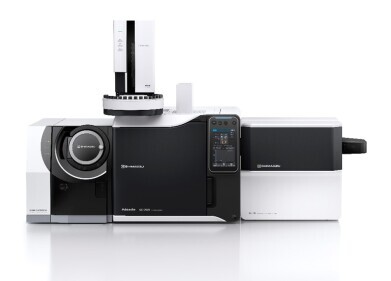
Advanced analytical tools like mass spectrometry have revolutionised the way research laboratories profile proteins. This protein characterisation technique is particularly useful for detecting the post-translational modifications (PTMs) that can change the behaviour of a protein. Mass spectrometry allows scientists to characterise these modifications in detail, with sophisticated instruments used to measure millions of spectra. Thousands of proteins and peptides can be identified in a single experiment, making it easier than ever to characterise post-translational modifications.
Orbitrap, a high-resolution mass spectrometry analyser manufactured by leading brands like Thermo Fisher, has become an invaluable tool for protein characterisation. Featuring both a central electrode and a pair of outer electrodes, the advanced instrument doubles as a detector and an analyser.
Mass spectrometers can be especially useful for detecting proteins using Peptide Mass Fingerprinting (PMF). The analytical technique separates the protein into smaller peptides, with a mass spectrometer used to measure the individual molecular weights of each peptide. Information is then entered into a computer program to compare the unique peptide mass combinations and identify the parent protein.
-
Quadrupole Time-of-Flight (QTOF) Mass Spectrometry
Quadrupole Time-of-Flight (QTOF) Mass Spectrometry is another high-resolution technique used to profile complex mixtures and characterise proteins. This advanced hybrid technique uses four parallel rods and a collision cell to enhance the capabilities of a conventional time-of-flight mass analyser. Mass-to-charge ratios are determined by forcing ions down the flight tube, with lighter ions reaching the detector faster. Impressive speeds and the capacity for quantitative analysis makes QTOF a staple in drug discovery laboratories. It’s also a highly regarded technique for clinical research, environmental screening, forensic toxicology and food safety applications.
-
Trapped Ion Mobility Spectrometry (TIMS)
Founded in ion mobility spectrometry (IMS) technology, TIMS is a gas-phase technique that allows researchers to capture a wide molecular weight range of signals. The technique separates ions according to mobility and utilises collision cross-section (CCS) data to allow researchers to get even more specific with protein analysis.
-
Dynamic Light Scattering (DLS)
Over the past decade, Dynamic Light Scattering has become a major asset for colloid characterisation. Fast and user friendly, the technique delivers results in just minutes without the need for high sample volumes. As well as characterising the size of the sample, DLS unlocks distribution information without the need for prior separation. This gives it an impressive range of applications, from low nanometer polymer solutions to high range particle dispersion.
-
Ultra High Performance Liquid Chromatography (UHPLC)
Fast and efficient, Ultra High Performance Liquid Chromatography uses pressure and speed to separate and analyse proteins. High-resolution results make it particularly valuable for evaluating the complex glycosylation patterns that can appear in proteins with post-translational modifications.
-
Centrifugation
Centrifugation is a mechanical process that separates proteins from a solution according to sedimentation rate. This is determined by factors such as shape, size and density, as well as the viscosity of the solution and speed of the centrifuge rotor. Centrifugal force is used to isolate proteins, which can then be individually analysed.
-
Gel Electrophoresis
Gel Electrophoresis relies on movement within an electric field to separate proteins according to molecular size. When exposed to the electric field, proteins are forced to unfold and are coated in a negative charge. This allows them to separate and individually gravitate towards the positively charged gel. From here, the proteins are transferred onto a membrane where they can be analysed using techniques such as Western blotting.
Also known as immunoblotting, the three-step method is used to isolate and identify individual proteins. Characterisation is achieved by flooding the membrane with an antibody solution specific to the target protein. This prompts molecular bands containing the target protein to bind to the antibody and change colour. For best results, it’s important to use high quality antibodies and calculate optimal concentrations before flooding the membrane.
-
Amino Acid Analysis
Protein characterisation is often complemented by Amino Acid Analysis techniques. This allows researchers to unlock even more insight into the unique characteristics of a sample. Laboratories utilise a variety of different chromatographic techniques to isolate, analyse and quantify amino acids. As well as mass spectrometry, these can include thin-layer chromatography, ion-exchange HPLC, paper chromatography, reversed-phase HPLC and capillary electrophoresis.
Protein characterisation in the biomedical sector
As well as supporting the human body, proteins are the building blocks of diseases. This makes protein characterisation critical to the biomedical research sector. Protein mapping is used to develop drugs and treatments, as well as create diagnostic reagents used to detect and screen for diseases.
Protein characterisation techniques have been fundamental to COVID-19 research, with advanced characterisation techniques used to understand more about the virus, as well as profile the aggressive spike protein that binds to the receptors of target cells and supports virus-cell fusion. This in-depth research helps arm scientist with the information they need to develop effective vaccines.
“The SARS-CoV-2 spike (S) protein serves as the most important target antigen for vaccine development based on preclinical research on candidate vaccines against SARS-CoV or Middle East respiratory syndrome coronavirus (MERS-CoV),” reads the study published in the Proceedings of the National Academy of Sciences of the USA. “The trimeric S protein is a prominent structure at the virion surface and essential for SARS-CoV-2 cell entry. As a class I viral fusion protein, it mediates virus interaction with the cellular receptor angiotensin-converting enzyme 2 (ACE2), and fusion with the host cell membrane, both key steps in infection. Thus, infection can be prevented by S-specific antibodies neutralizing the virus.”
Want to know more about the role protein characterisation plays in the biomedical research field? Dr Zhongyi Cheng spotlights the benefits of combining modern Trapped Ion Mobility Spectrometry (TIMS) with Time-of-Flight Mass Spectrometry (TOF-MS) in ‘Deep characterisation and quantitative analysis of proteins and post-translational modifications - How TIMS has extended the capabilities of MS and shown considerable potential for improving PTM identification and critical understanding of sign.'
Digital Edition
Lab Asia 31.2 April 2024
April 2024
In This Edition Chromatography Articles - Approaches to troubleshooting an SPE method for the analysis of oligonucleotides (pt i) - High-precision liquid flow processes demand full fluidic c...
View all digital editions
Events
Apr 28 2024 Montreal, Quebec, Canada
May 05 2024 Seville, Spain
InformEx Zone at CPhl North America
May 07 2024 Pennsylvania, PA, USA
May 14 2024 Oklahoma City, OK, USA
May 15 2024 Birmingham, UK


