Microscopy & microtechniques
Published over 5 years ago. See the latest and most current information on Microscopy & microtechniques.
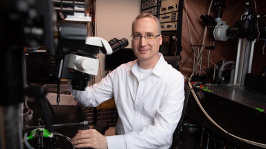
A team of scientists from the University of Southern California (USC) has uncovered an exciting new use for tattoo ink, with lead researcher Cristina Zavaleta asserting it could be used to detect cancerous cells. Using the same high pigment dyes found in tattoo studios around the world, Zavaleta and her team developed innovative imaging contrast agents that bind to nanoparticles and illuminate cancer cells.
The agents were developed at the USC Viterbi Department of Biomedical Engineering, with the findings published in the journal Biomaterials Science. With more than 38% of Americans affected by some form of cancer during their lifetime, the research could have incredible implications for public health. Survival rates are exponentially higher in patients where cancer cells are detected early, making the new colour-based imaging agents a hugely valuable diagnostic tool.
To create the imaging agents, the team infused cancer-targeting nanoparticles with the same FDA-approved dyes found in tattoo ink. When injected into a patient these optical inks enhance the visibility of nanoparticles and make it easier for doctors to detect cancerous cells during MRI and CT scans. The use of colour-enhanced imaging agents also makes it easier for surgeons to remove every part of a tumour and reduce the risk of future growth.
"For instance, if the problem is colon cancer, this is detected via endoscopy," explains Zavaleta. "But an endoscope is literally just a flashlight on the end of a stick, so it will only give information about the structure of the colon - you can see a polyp and know you need to take a biopsy. But if we could provide imaging tools to help doctors see whether that particular polyp is cancerous or just benign, maybe they don't even need to take it," she adds.
Thinking outside the box, Zavaleta drew inspiration from a digital animation class she attended in California. She was instantly drawn to the brightly coloured inks and paints used by the artists and wondered if their properties could be transferred to a medical context.
“I was thinking about how these really high pigment paints, like gouache watercolours, were bright in a way I hadn't seen before, and I was wondering if they had interesting optical properties,” says Zavaleta. "I remember I brought a 96-well plate and he squirted tattoo ink into each of the wells. Then I took the inks to our Raman scanner (used to sensitively detect our tumour-targeting nanoparticles) and discovered these really amazing spectral fingerprints that we could use to barcode our nanoparticles. It was super cool."
Want to know more about the latest imaging technologies? Don’t miss ‘The intracellular world in 3D and in colour: developments at the biological cryo-imaging beamline B24 at the UK’s national synchrotron, Diamond Light Source.’
ILM 51.5 July 2026


-(1).jpg)
.jpg)