Microscopy & microtechniques
Researchers have reviewed the technical bottlenecks that still limit intravital mesoscale microscopy, despite rapid progress in optical hardware and computational methods. The analysis sets out how instrument designers and biologists must trade off resolution, imaging speed and field of view while also seeking to limit light scattering, optical aberrations and photodamage in living tissue
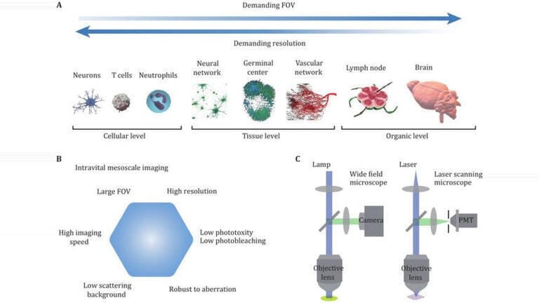
Intravital mesoscale imaging – made from within living tissue – has played an increasingly important role in life-sciences research because it can connect observations at cellular scale with measurements that capture organ-level organisation and physiology. By allowing researchers to image living tissue across comparatively large fields of view while still resolving fine structure, mesoscale approaches have offered a practical route to study how local cellular events relate to broader function, from microvascular dynamics to tissue remodelling.
A recent review has assessed the persistent technical constraints that continue to shape what intravital mesoscale optical imaging can achieve, even as optical microscopy has improved markedly. The authors described intravital mesoscale imaging as a problem of optimisation rather than a single target specification.
In practice, researchers must choose an operating point that balances spatial resolution, acquisition speed and field of view, because improvements in one domain often impose penalties elsewhere. These trade-offs become particularly acute in vivo, where motion, optical heterogeneity and tight limits on permissible light exposure can reduce image quality and restrict experimental duration.
The review identified several fundamental obstacles that remain difficult to remove. Light scattering in tissue can degrade contrast and restrict imaging depth, particularly beyond superficial layers. Optical aberrations, whether introduced by the microscope or by the sample itself, can distort the wavefront and reduce effective resolution across wide fields. Phototoxicity and photobleaching continue to constrain longitudinal experiments, because repeated excitation can perturb biological processes and deplete fluorescent signal.
The authors noted that these factors do not act in isolation. Higher excitation intensity can improve signal and support faster imaging but it can also increase photodamage, while attempts to image deeper structures can require optical strategies that reduce speed or complicate instrumentation.
Against this background, the review surveyed the principal families of optical and computational approaches that research groups have used to extend intravital mesoscale performance. Wide-field methods can provide rapid acquisition across large areas, which can suit dynamic processes that unfold quickly, but they can struggle to maintain optical sectioning and contrast in scattering samples.
Laser-scanning strategies can improve sectioning and image quality but they typically trade speed and field of view against resolution and signal-to-noise, particularly when experiments demand volumetric imaging. The authors also highlighted the growing role of computational imaging, in which reconstruction, denoising, deconvolution or model-based inference can recover information that the raw optical signal does not directly provide, or can help to correct distortions that arise in vivo.
The review concluded that further gains would depend on coordinated progress in optics, fluorophore chemistry and computation. The authors argued that future development is likely to prioritise deeper imaging in living tissue, alongside improvements in the space–bandwidth product, which captures how much spatial information an imaging system can acquire across a field of view.
They also pointed to the need to integrate computational methods more tightly with image acquisition, with an emphasis on real-time processing and analysis pipelines that can handle large datasets during experiments rather than after them. Such integration, the review suggested, could allow researchers to adapt acquisition on the fly, manage data volumes more effectively and extract biologically relevant signals at scale, which could broaden the reach of intravital mesoscale imaging across physiology, immunology, neuroscience and other fields.
For further reading please visit: 10.52601/bpr.2025.250015
ILM Guide 2026/27