-
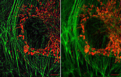 Mitochondria: Copyright MRC LMB/MBU* Left shows structured illumination microscopy (SIM) super resolution image of actin fibres (green) and mitochondria (red) in bovine pulmonary artery endothelial cells. Stained with MitoTracker Red CMXRos for labeling mitochondria, Alexa Fluor 488 phalloidin for labeling F-actin. Scale bar 5 microns. The image on the left shows the same sample as a conventional wide field fluorescence image. SIM super resolution resolution by a factor of about 2 in X, Y and Z. *LMB/MBU = MRC Laboratory of Molecular Biology/Mitochondrial Biology Unit
Microscopy & Microtechniques
Strathclyde playing a crucial role in microscope revolution
Mar 04 2013
Biomedical research across the UK – including a pioneering University of Strathclyde project – is to benefit from a £25.5 million cash injection to boost the resolution revolution in microscope technology.
Three of the UK's research councils - the Medical Research Council (MRC), the Biotechnology and Biological Sciences Research Council and the Engineering and Physical Sciences Research Council – have invested £20.1 million, £2.4 million and £2 million respectively, to establish 17 microscopy platforms that will bring about ground-breaking advances in biological and biomedical research.
These include a £1.7 million MRC award to a Strathclyde team led by Professor Gail McConnell, of the Centre for Biophotonics, to develop a prototype of a unique lens capable of producing stunning, laser-scanned 3D images of disease tissues, with sufficient detail to see inside individual cells.
Professor Steve Hill, who chaired the expert panel which assessed the proposals, said: “Microscopy is one of the most important tools scientists have for discovery-based research but the high costs associated with this technology are often a barrier to expansion. This funding is crucial to help the UK capitalise on the latest technologies and maintain its internationally leading position in biological and biomedical research.
”This type of microscopy relies on scientists in very different disciplines coming together to solve very specific imaging problems. All 17 projects were able to demonstrate extremely strong partnerships between biologists, physicists, chemists, mathematicians, engineers, technologists and equipment manufacturers."
Digital Edition
Lab Asia 31.2 April 2024
April 2024
In This Edition Chromatography Articles - Approaches to troubleshooting an SPE method for the analysis of oligonucleotides (pt i) - High-precision liquid flow processes demand full fluidic c...
View all digital editions


.jpg)


Events
May 21 2024 Lagos, Nigeria
May 22 2024 Basel, Switzerland
Scientific Laboratory Show & Conference 2024
May 22 2024 Nottingham, UK
May 23 2024 Beijing, China
May 28 2024 Tel Aviv, Israel


