News
Expansion of imaging infrastructure nationwide will ramp up opportunities for both faster patient diagnosis and investigational drug development
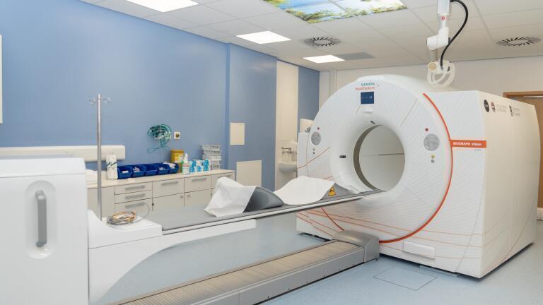
Scotland’s first Positron Emission Tomography (PET) total-body scanner has entered service at the Royal Infirmary of Edinburgh. Delivered by the National PET Imaging Platform (NPIP), the scanner will be co-managed by the universities of Edinburgh and Glasgow. NPIP is funded by a £32 million investment from the UKRI Infrastructure Fund and is delivered in collaboration by the Medical Research Council, Innovate UK and Medicines Discovery Catapult.
The Biograph Vision Quadra PET/CT scanners – now operational in London and Edinburgh – are manufactured by Siemens Healthineers, which also provides on-site scientific staff to support day-to-day operations at both sites. The London NPIP scanner, located at St Thomas’ Hospital, was formally inaugurated on 27 November 2024. The first patients received scans shortly thereafter, making it the first government-funded scanner in the NPIP programme to become operational. It is jointly managed by Imperial College London and King’s College London. A third scanner, situated at the Royal Free Hospital in London, sits outside the UKRI scheme, having been funded by the Royal Free Charity. The Royal Free's full-body scanner was the first PET scanner to be operational in the UK, receiving its first patients from March 2024.
The installation of these machines significantly enhances the NHS’ capacity to diagnose and treat cancer, cardiovascular, inflammatory and other diseases. The network consolidates the UK’s global leadership in PET technology, which is capable of accelerating diagnosis and drug development, ultimately improving patient outcomes. The integration of these scanners establishes a critical and clinical national infrastructure that facilitates collaboration between clinicians, industry and researchers, attracting pharmaceutical trials to the UK.
Total-body PET scanners offer a step change in capability: they are up to 40 times more sensitive, up to 10 times faster and can scan 50% more patients daily compared with previous-generation systems.
“The national platform we have created allows the combined power of technology and data to be harnessed, attracting industry to test their new treatments here in the UK for the benefit of our patients and our economy,” said Professor Chris Molloy, Chief Executive of Medicines Discovery Catapult, one of the NPIP partner organisations.
“It shows what is possible when strategic public funding, clinical expertise, industry knowledge and academic excellence come together around a shared national purpose. These revolutionary scanners help save lives and create large-scale capability for radiopharmaceuticals and AI-enabling datasets,” he added.
Dr Juliana Maynard, Director of Operations and Engagement at NPIP, said: “We see the NPIP network as both critical and clinical national infrastructure. It is a connected nationwide network for data sharing, discovery and innovation that we could only have imagined a decade ago.
“Using these total-body PET scanners, we can observe disease in real time across the entire body and now, across the entire country. That is game-changing for drug discovery and treatment in the UK and, more importantly, for how quickly patients can benefit from it.
“Researchers will gain access to vastly improved clinical data, not only by tapping into the network for their own trials, but from every study connected to the platform. This will create an unprecedented level of collaboration in imaging, placing the UK firmly on the world map as a centre of excellence,” she said.
The Medicines Discovery Catapult-led PET scanner network enables a transformative imaging ecosystem. It enhances biological insight, expedites translational medicine and ensures equitable access to leading-edge clinical research tools. This represents a key element in the strategic expansion of UK drug discovery infrastructure.
PET is a powerful imaging modality that allows clinicians and researchers to observe biological processes at the molecular level and in near real time. Unlike structural imaging techniques such as X-ray or computed tomography (CT), which show anatomical detail, PET captures physiological and biochemical activity. This marks a substantial advance in medical imaging, particularly within oncology, neurology and cardiology.
PET scanners function by detecting gamma rays emitted indirectly by a positron-emitting radionuclide (tracer) introduced into the body. The most widely used tracer is fluorodeoxyglucose (FDG), a glucose analogue labelled with the radioactive isotope fluorine-18. Administered intravenously, FDG accumulates in tissues based on their metabolic activity. For example, highly active tissues such as cancer cells absorb more FDG.
Fluorine-18 undergoes radioactive decay and emits a positron. This positron travels a short distance before encountering an electron, producing an annihilation event that generates two gamma photons travelling in opposite directions. The PET scanner detects these photons using a ring of detectors around the patient and triangulates their origin through coincidence detection. By reconstructing millions of such events, the scanner creates a three-dimensional image showing the tracer’s spatial distribution.
With a short half-life of approximately 109 minutes, fluorine-18 is well suited to PET imaging, particularly where rapid pharmacokinetics are desirable.
PET represents a significant advance over traditional imaging techniques such as X-ray, ultrasound and CT by offering functional rather than purely anatomical information. Key advantages include:
Strategically, the NPIP programme is expected to accelerate drug discovery by providing high-quality longitudinal data on investigational new drugs, including information on distribution, target engagement and physiological effects.
Access to near real-time pharmacokinetic data will support timely go or no-go decisions in R&D across multiple disease areas including oncology, neurology, cardiovascular disease and paediatrics. A national PET data repository will be established to aggregate imaging data and provide an openly available resource to benefit academia, industry and patient care.
The first PET scanner for human studies was installed at the University of Pennsylvania in the early 1970s. The clinical development of PET is closely associated with American physicist Michael Phelps, who built on pioneering work conducted at the Mallinckrodt Institute of Radiology at Washington University in St Louis, led by Michel Ter-Pogossian.
ILM Guide 2026/27


.jpg)


2.jpg)
