

News
An ELRIG webinar has explored how cell-based screening supports drug discovery, from early target validation to advanced three-dimensional disease models, with speakers outlining the trade-offs between throughput, complexity and physiological relevance
ELRIG’s recent webinar ‘Cell-Based Screening 101’, delivered online in April 2026, has offered a detailed introduction to the role of cell-based assays in contemporary drug discovery, with contributions from scientists at AstraZeneca, Domainex and other organisations active in the field. The session has examined what constitutes a cell-based assay, where such assays sit within the drug discovery pipeline, and why researchers must balance throughput, model complexity and physiological relevance when they select an experimental system.
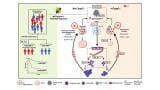
At the centre of the discussion was the idea that cell-based assays occupy a critical middle ground in biomedical research. In contrast to biochemical or biophysical assays, which rely on purified proteins and simplified systems, cell-based assays allow researchers to observe how compounds – or genetic interventions – affect living cells in a functional context. At the same time, they remain more controllable and scalable than animal studies. This combination has established them as a cornerstone of early-stage drug discovery and an increasingly influential component of later development and regulatory decision-making.
A cell-based assay typically begins with the culture of living cells in vitro, followed by deliberate perturbation and measurement of a biological response. Perturbations may include exposure to small molecules, targeted genetic modification such as gene knockout, or other interventions designed to alter cell behaviour. The defining characteristic is that the readout reflects a cellular event rather than a simple molecular interaction. Such readouts may include changes in protein expression, cell morphology, extracellular matrix deposition, gene expression or viability, as well as more complex phenotypic outcomes.
The speakers placed these assays within the broader context of drug discovery, which they described as a (potentially) long, costly and resource-intensive process. It may take more than a decade from target conception of an investigative drug to its regulatory approval. Within this timeline, cell-based assays have traditionally been most prominent in the preclinical phase, including target identification, validation, hit identification, lead optimisation and candidate selection. Their importance at the outset lies in the ability to establish whether a proposed target has a genuine link to disease biology before further investment proceeds.
During hit identification, researchers may screen large compound libraries to identify molecules that influence a target of interest. At this stage, assay selection depends on both biological relevance and practical constraints. Biochemical, biophysical and cell-based approaches may all be used. As programmes progress and candidate molecules emerge, cell-based assays become more central because they allow confirmation that compounds produce a meaningful functional effect within a cellular system rather than simply bind to a molecular target.
A major theme throughout the seminar was the balance between simplicity and biological realism. Large screening campaigns, which may involve potentially millions of compounds, typically rely on simpler, more scalable models. Immortalised cell lines – such as HeLa – are widely used in this context because they proliferate indefinitely, are cost-effective and adapt readily to automation. However, repeated culture and immortalisation may alter their biology, which means they do not always reflect the behaviour of native tissue.
Primary cells provide greater physiological relevance because they are derived directly from tissues or donors. They more closely resemble native biology but are more difficult to culture, less scalable and subject to variability between donors – as well as adding cost to the project. Their limited lifespan in vitro adds further constraints. The webinar also addressed induced pluripotent stem cells, which offer a route to generate specialised cell types such as neurons or cardiomyocytes. These systems can bridge some of the gap between scalability and relevance, although they remain technically demanding and expensive.
The discussion then turned to assay formats and recognised that two-dimensional monolayer cultures remain the simplest and most widely used system, with cells grown on plastic surfaces in flasks or multiwell plates. This format allows straightforward compound exposure and supports a wide range of readouts. The webinar emphasised that such models – although simple – can still provide substantial insight when designed carefully. A fibrosis assay described as a ‘scar-in-a-jar’ model illustrated this point. By monitoring extracellular matrix deposition and myofibroblast activation in response to transforming growth factor beta and therapeutic intervention, researchers can observe dose-dependent changes in collagen production and related markers.
Successful screening depends not only on biological design but also on time, cost and compatibility with automation. The guiding principle presented was to maintain simplicity wherever possible while retaining sufficient complexity to answer the biological question. Greater complexity reduces throughput (time) but increases operational demands. Robotic systems, automated liquid handling and miniaturised plate formats such as 1536-well plates allow the rapid processing of large compound libraries, although they impose practical limits on assay design.
The seminar also highlighted cell painting as an example of a data-rich phenotypic screening approach. This method uses fluorescent dyes to label cellular structures and organelles, followed by high-resolution imaging and computational analysis to extract morphological features. These features create a phenotypic fingerprint that allows comparison across known compounds and their features. Researchers can use such comparisons to infer mechanism of action or to identify compounds with toxicity profiles similar to known reference molecules.
As drug discovery progresses and the number of candidate compounds decreases, more complex models can be introduced. The webinar described a progression from two-dimensional cultures to three-dimensional spheroids, organoids and microphysiological systems such as organ-on-a-chip platforms. These models better replicate tissue organisation and microenvironmental conditions, including gradients of nutrients and oxygen. For example, in tumour organoids cells may develop proliferative, quiescent and necrotic zones, each with distinct biological characteristics.
The quiescent zone received particular attention because it may contain cells that resist treatment and later drive disease recurrence. More complex models can reveal such features, which may not appear in simpler systems. However, increased physiological relevance comes with reduced throughput, higher cost and greater variability.
Microphysiological systems extend this complexity further by introducing elements such as fluid flow and mechanical forces, which influence drug distribution and cellular response. These systems may offer improved translation from in vitro findings to in vivo outcomes, but they are not suited to large-scale screening and are typically reserved for later-stage studies.
A key message from the webinar was that no single model provides a complete representation of biology. Assay selection must begin with a clear definition of the biological question. Multiple models may be required across a project, each chosen for its ability to address a specific aspect of the problem. The value of an assay lies in its ability to produce reliable and interpretable data rather than in its apparent complexity.
The discussion also reflected broader changes in the regulatory landscape. Cell-based and other in vitro approaches have increasingly replaced animal studies in both efficacy and toxicology testing. Regulatory attitudes have begun to shift, with recent policy changes in the USA removing mandatory animal testing requirements – and starting down the road to phasing them out altogether – while also encouraging submission of data already from advanced cell-based models. Funding initiatives to support organoid standardisation have reinforced this direction.
Overall, the seminar presented cell-based assays as an essential and expanding component of drug discovery. It showed that these approaches span a continuum of models, from highly scalable monolayers to complex tissue-like systems. Their strength lies in the ability to connect molecular activity with functional biological outcomes, and their importance is likely to increase as the field seeks more predictive and ethically acceptable approaches to therapeutic development.
Summary of key takeaways
ILM Guide 2026/27


2.jpg)
