News
Published over 10 years ago. See the latest and most current information on News.
Researchers from the National Physical Laboratory (NPL), UCL and the Royal Free Hospital have differentiated between patients with a rare bleeding disorder and healthy volunteers using super-resolution microscopy, providing an alternative method for accurately and cost-effectively diagnosing rare platelet diseases.
Platelets form part of the blood and they help heal wounds and prevent bleeding by forming blood clots. They do this through tiny granules which release molecules for blood clotting. Platelet disorders occur when these granules are too few in number, are misshapen or don't release the right molecules. As the causes for platelet disorders vary so much, specific treatment can be improved if diagnostic tests can distinguish the different type
Microscopy provides an excellent diagnostic approach, but to study tiny platelet granules, electron microscopy (EM) is necessary. This is costly, requires fresh samples, gives limited information about what is wrong and is not widely available in the NHS for patients with bleeding disorders. As such, many patients are 'over-treated' for platelet disorders through blood transfusions and other invasive treatments, which can be high-risk for the patient, as well as resource-intensive for the healthcare system.
First author and MRC PhD student David Westmoreland, UCL Life Sciences, said: "We've found that SIM has a lot of advantages over whole mount electron microscopy as a diagnosis method. Samples don't need to be analysed live and can be reanalysed, and automation means analysis is unbiased and less time-consuming. Given about 75% of patients with a bleeding disorder such as Hermansky-Pudlak Syndrome are initially misdiagnosed, and 28% need to see between four to six specialists before receiving the correct diagnosis, there is a demand for a new method of analysis."
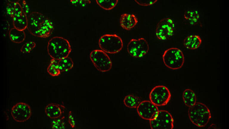
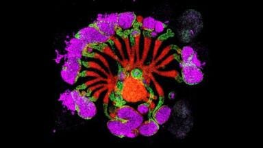
For the proof-of-concept study, the team used Structured Illumination Microscopy (SIM) to specifically image platelet granules in blood samples using a marker protein, CD63. The imaging technology was custom-built by the team to automatically count the number of granules per platelet, identifying those with Hermansky-Pudlak Syndrome, a rare blood disorder thought to affect 1 in 500,000.
The team distinguished the three patients with Hermansky-Pudlak Syndrome from the seven normal controls with 99% confidence. Automated counting of granules showed that those with the disorder had only one third as many granules as controls.
Corresponding author, Professor Dan Cutler, MRC Laboratory for Molecular Cell Biology at UCL, said: "Our limited analysis of this new method is extremely promising and we hope to take it forward to test for multiple parameters with different markers. In this way, we could use a single super-resolution image to screen for many different platelet-based blood disorders."
Alex Knight, Principal Research Scientist, in the Biotechnology Group at NPL, said "The results of the SIM imaging were very dramatic and a significant improvement over what we see with conventional imaging. The accuracy of the results have the potential to translate into a much more viable and accessible means of identifying blood disorders. Super-resolution microscopies have contributed to many of the breakthroughs in life sciences research in recent years, even winning the Nobel Prize for Chemistry in 2014. This study, however, shows the significant impact the technique could have in applications outside of research too."
Keith Gomez, Senior Lecturer in Haemostasis, Haemophilia Centre and Thrombosis Unit, Royal Free Campus, UCL Medical School, said: "For clinicians struggling to access electron microscopy facilities, SIM offers an easy to use and very informative alternative. This new technique has the potential to make diagnosis of structural platelet disorders much easier for clinical staff in the NHS which will lead to better patient care."
Funding for the work was kindly provided by the Medical Research Council (MRC), the NMS Chemical and Biological Metrology programme and A*STAR (Agency for Science, Technology and Research, Singapore).
Results of the study will be published in the Journal of Thrombosis and Haemostasis
ILM Guide 2026/27


.jpg)