News
Published over 7 years ago. See the latest and most current information on News.
Report by Helen Miller from the University of Oxford
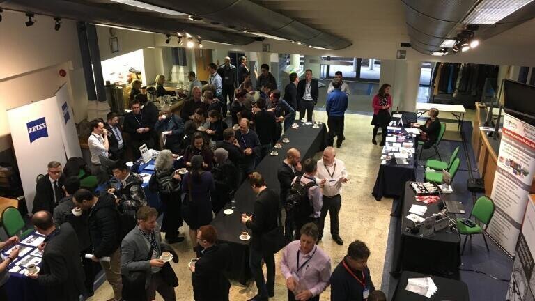
The Royal Microscopical Society’s Frontiers In BioImaging conference was held at the Technology and Innovation Centre at the University of Strathclyde between June 27-28th 2018, moving out of London for the first time. The meeting was well attended with enthusiastic microscopists eager to discuss the latest developments such as technological breakthroughs and addressing important biological questions with microscopy.
The programme consisted of six packed sessions including seven invited talks and four flash talks from industry, an industry exhibition, a poster session and an excellent conference dinner. The conference started with a session including a talk on Brillouin Microspectroscopy from Francesca Palombo at the University of Exeter. Brillouin Microspectroscopy is a technique that finds material properties such as the elastic modulus and refractive index from optical measurements.
Post lunch we were treated to some technological and methodological developments, ranging from Graphene Oxide films as long-lifetime contrast enhancement factors, diamond embedded electrospun nanofibers for investigating mechanical properties and light sheet imaging of the far-from-coverslip slide of T-cells combined with 3D imaging using a double-helix point spread function. An unfortunate illness meant a space in the programme was excellently filled with a talk on the future of the Mesolens including wonderful images of whole flies in 3D.
I presented my own work on electroporating proteins labelled with fluorescent tags into bacteria for 3D single molecule tracking on the second day. This method allows exquisite control over the number of labelled proteins introduced to a cell, meaning long-lived fluorescent probes can be used for tracking in low background environments. After my talk I had lots of stimulating conversations with people about how the technique could be applied to the biological questions they are working on and some excellent suggestions for software I could consider using.
As always at this meeting, the many conversations with other researchers have given me lots to think about as I return to the lab bench with renewed enthusiasm. I would like to thank the Royal Microscopical Society for their generous financial support which enabled me to attend this meeting.
ILM 51.5 July 2026


-(1).jpg)
.jpg)