Laboratory products
Published over 12 years ago. See the latest and most current information on Laboratory products.
The risks associated with radiation contact in children could be reduced thanks to a new method of performing scans. The BBC reports that a new way of scanning children's livers for tumours could cut down on how much unnecessary radiation they are exposed to.
Exposure to radiation in childhood can increase the likelihood of getting cancer in later life. Doctors in London have developed a new method for screening liver tumours without providing children with a high dose of radiation, reports the news provider.
Doctors at King's College Hospital are currently trialling the new technology that could help to locate tumours in an easier and safer way. The study has been published in the 'European Journal of Ultrasound'.
Ultrasound scans can be used to detect lesions on nodules on the livers of children that are suffering from liver disease. The process can also identify these problems in youngsters that have fatty livers. However, the ultrasound is not able to tell whether these lesions are benign, which would mean that they can be disregarded, or malignant, which need fast treatment, reports the news provider.
In order to make this differentiation a CT scan can be used, however, this subjects the child to high doses of radiation, which could be damaging to health in the future. The new method was developed as a way of reducing unnecessary exposure, while still giving a definitive diagnosis.
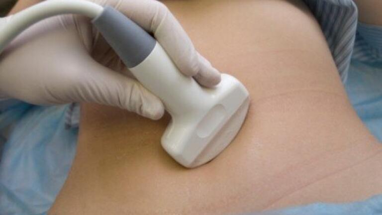
Doctors are now testing a version of ultrasound that has been upgraded to give a clearer picture. This type of ultrasound has been used in adults for a number of years, but has not yet been used in children, according to the BBC.
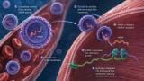
It works by injecting a chemical into the arm, which is entirely harmless and has no effect on the patient. The chemical then forms microscopic bubbles throughout the bloodstream, which serves as a "contrast agent" during the ultrasound.
Professor Paul Sidhu told the BBC that the chemical means the arteries light up, which then spreads to the veins and ultimately the entire liver. He described the effect as making the liver look like a "field of gold". In comparison to the healthy liver, any cancerous tumour will quickly dissipate the chemical, which will make it appear as a dark area on the scan.
A trial of 44 children, all suffering with chronic liver problems, found that this new method was able to provide an accurate diagnosis for each patient, reports the news provider.
ILM 51.5 July 2026


-(1).jpg)
.jpg)
.jpg)