Research news
A new imaging approach developed through a collaboration between the University of Oxford and ZEISS is entering a proof-of-principle phase, with the potential to reshape how drug behaviour is studied at the cellular level.
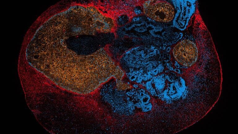
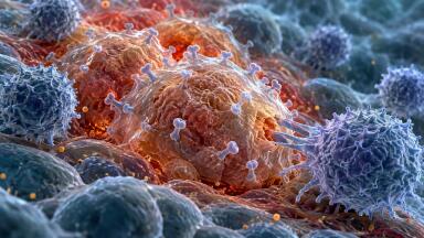
At the centre of the work is Selective Plane Illumination Lattice Light Sheet Microscopy (SPI LLSM), an advanced technique designed to track how drugs move and interact within complex biological systems. By using ultrathin, non-diffracting light sheets, the method enables high-resolution visualisation of drug dynamics in three-dimensional models, including organoids and tissue biopsies.
The technology has been developed to address limitations in existing approaches such as fluorescence correlation spectroscopy, offering a faster and more consistent way to quantify drug interactions in biologically relevant environments. Earlier studies demonstrated its capability in solution assays and cell cultures, and the next phase will extend its use to more complex systems, including patient-derived tissues.
“This innovation enables observation of 3D reaction–diffusion dynamics in organoids and tissue biopsies, with potential to transform drug screening,” said Professor Marco Fritzsche, Scientific Director of the Oxford-ZEISS CoE.
Over the next 12 months, researchers from academia and industry will work alongside ZEISS R&D to evaluate the system’s performance in drug profiling applications. The aim is to determine whether SPI LLSM can deliver reliable, scalable insights for drug discovery, particularly in areas such as cancer and autoimmune disease.
By enabling direct observation of reaction–diffusion processes in intact 3D models, the approach opens new possibilities for understanding how therapeutics behave in realistic biological contexts. If successful, it could offer an alternative to conventional imaging methods and support more predictive drug screening workflows.
The results of the study will guide further development and potential commercialisation, with the longer-term aim of making the technology broadly accessible for research and pharmaceutical applications.
More information online
ILM 51.6 Sept 2026



 Product directory












,-photo-Karin-Allander.jpg)




.jpg)