Wellcome Image Awards 2011
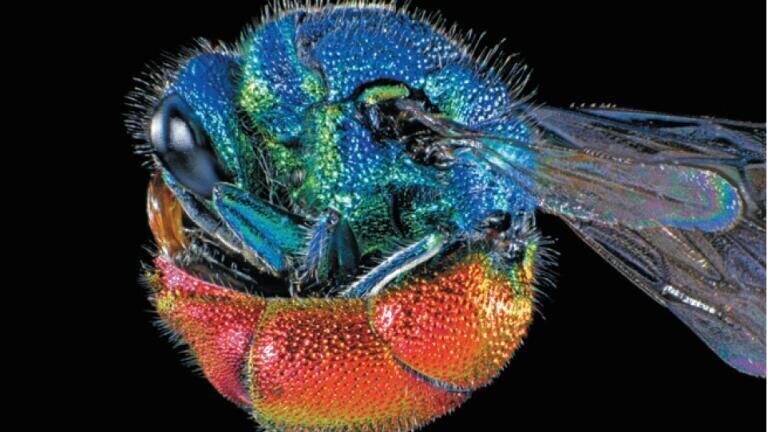
Ruby-tailed wasp. Spike Walker/Wellcome Images. This photomicrograph shows an adult ruby-tailed wasp curled into a ball. Chrysis ignita is the most commonly observed of several species of the ruby-tailed wasp. The wasp was lit with two electronic flashes while imaging to highlight the natural iridescent colours on its body.
Moth wing scales. Kevin Mackenzie, University of
Aberdeen/Wellcome Images. Scanning electron
micrograph (SEM) of the scales on the wing of a
Madagascan moon moth, Argema mittrei. This moth is
also known as the Comet moth, after its very long tail.
The tail span is 15 cm and wing span 20 cm, making it
one of the world\'s largest silk moths.
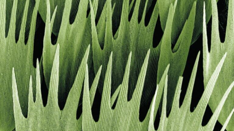
Moth wing scales. Kevin Mackenzie, University of Aberdeen/Wellcome Images. Scanning electron micrograph (SEM) of the scales on the wing of a Madagascan moon moth, Argema mittrei. This moth is also known as the Comet moth, after its very long tail. The tail span is 15 cm and wing span 20 cm, making it one of the world\'s largest silk moths.
Pyramidal neurons. Michael Häusser and Hermann Cuntz, UCL/Wellcome Images
This computer simulated image shows synthetic pyramidal neurons, of optimised size,
shape and connectivity, that are
indistinguishable from those found in the real biological brain. Pyramidal cells are so-called as
they have a pyramid-shaped cell body (soma), and are also characterised by long branching
dendrites. They are found in the forebrain (cortex and hippocampus) of mammals and are thought to be involved in cognitive function.
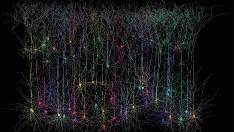
Pyramidal neurons. Michael Häusser and Hermann Cuntz, UCL/Wellcome Images This computer simulated image shows synthetic pyramidal neurons, of optimised size, shape and connectivity, that are indistinguishable from those found in the real biological brain. Pyramidal cells are so-called as they have a pyramid-shaped cell body (soma), and are also characterised by long branching dendrites. They are found in the forebrain (cortex and hippocampus) of mammals and are thought to be involved in cognitive function.

Microscopy & microtechniques

Wellcome Image Awards 2011

18 Apr, 2011

Published over 15 years ago. See the latest and most current information on Microscopy & microtechniques.

Winners of the Wellcome Image Awards 2011, (presented 23rd February) have used techniques ranging from clinical photography to brain tractography to capture the wonder of medicine and the life sciences in all of their glorious splendour. The selected images are on display at Wellcome Collection until July 2011. Each image is accompanied by ’the story behind the picture’, to explain how it was created, what it adds to scientific understanding and why the judges felt it stands out. Selected images can also be seen on the website, Wellcome Images. Catherine Draycott, Head of  Wellcome Images, added: "It’s easy to forget that behind each of these intriguing images is a complex story of scientific pursuit. We search out hundreds of images from working scientists and artists each year and the awards offer a superb opportunity for people to find out about the cutting-edge science and the people behind the pictures."

Latest News

Lab Asia 33.4 - August 2026

Explore our Digital Edition

Discover the latest news and research

Digital edition

Explore Our Other Sites

Envirotech Online
Designed for semiconductor. Proven across industries.
Explore more Arrow
Pollution Solutions Online
Oil pipeline decommissioning in Wales secures environmental safety for future generations
Explore more Arrow
Petro Online
Flash point testing aligned with petrochemical compliance requirements
Explore more Arrow
Chromatography Today
Unlock high-resolution analysis of therapeutic oligonucleotides
Explore more Arrow