
Microscopy & Microtechniques
Major investment in new microscope technologies
Mar 04 2013
Major investment in new microscope technologies
A project led by the University of York and the Cancer Research UK London Research Institute (CRUK LRI), aims to combine light and electron microscopes into a single system to analyse how cells and tissues change during disease and infection.
It has been allocated £1 million from the Medical Research Council (MRC), the Biotechnology and Biological Sciences Research Council (BBSRC) and the Engineering and Physical Sciences Research Council (EPSRC) as part of the current funding round of the cross-Council Next Generation Optical Microscopy initiative launched in May 2012. Additional investment from the University of York, CRUK and commercial collaborators is expected to raise the funding to £2.million.
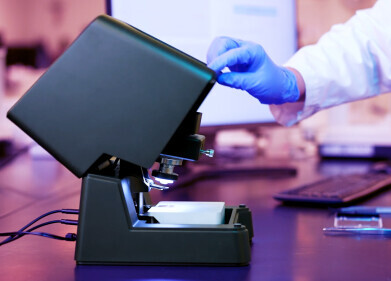
The new light microscope combined with an electron microscope being developed at York is the only one of its kind in Europe. Applications include imaging of subcellular processes related to cancer, better understanding of tumour biology and cancer cell invasion, and new insights into neurodegenerative diseases.
The project is led by Dr Peter O’Toole of the Imaging & Cytometry Laboratory in York’s Department of Biology, in collaboration with Dr Lucy Collinson of the Electron Microscopy Unit at CRUK LRI. Also involved are instrument manufacturers JEOL and DELMIC.
Dr O’Toole said: “Currently light microscopes allow us to watch real time events in cells and tissues so that we can understand basic biological functions and the changes that occur in disease and infection. Electron microscopes have taught us much about the fine details of cellular structures thanks to their fantastic resolution, but living material cannot be readily imaged and must be 'fixed' to halt the processes of life.
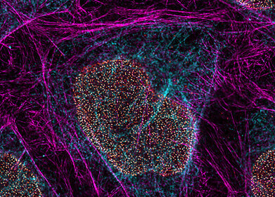
“Our approach is based on exploiting new ways of preparing cells and tissues, so that they can be seen simultaneously using light and electrons. This novel project will now combine the two microscopes to produce more informative images and help solve a multitude of biomedical questions.”
Dr Richard Treisman, Director of the Cancer Research UK London Research Institute, said: “This exciting new initiative to combine light and electron microscopy in 3D will open a new window into how cells and tissues function in health and disease."
Digital Edition
Lab Asia 31.2 April 2024
April 2024
In This Edition Chromatography Articles - Approaches to troubleshooting an SPE method for the analysis of oligonucleotides (pt i) - High-precision liquid flow processes demand full fluidic c...
View all digital editions


.jpg)


Events
May 21 2024 Lagos, Nigeria
May 22 2024 Basel, Switzerland
Scientific Laboratory Show & Conference 2024
May 22 2024 Nottingham, UK
May 23 2024 Beijing, China
May 28 2024 Tel Aviv, Israel


