News
Published over 8 years ago. See the latest and most current information on News.
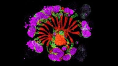
Researchers at the Crick have recently launched a biomedical project, Etch a Cell, which is using citizen science to identify features of interest in order to construct 3D models of the area under study. Individual and collaborative research using advanced EM microscopes available at the Crick, generates a huge number of images of molecules, cells and tissues which can be used to help understand cancer, infectious diseases (including HIV, tuberculosis, malaria), the immune system, the brain and nervous system, diabetes and more. To extract meaning from the image data produced, the Crick is appealing to individuals to help segment, or to draw around the cell features of interest.
In this project, the feature of interest is the nuclear envelope, the barrier that seperates the genetic information (DNA) inside the nucleus from the chemicals and reactions going on in the rest of the cell. By segmenting many images it is possible to create a 3D model of the nuclear envelope, allowing healthy and diseased cells to be studied in great detail.
Dr Martin Jones, a researcher at The Crick explained “Our project, Etch a Cell is designed to allow citizen scientists to draw segmentations directly onto our images in the Zooniverse web interface. The first task we have set is to mark the nuclear envelope that separates the nucleus from the rest of the cell – a vital structure where defects can cause serious problems. These segmentations are extremely useful in their own right for helping us understand the structures, but citizen science offers something beyond the already lofty goal of matching the output of an expert. By allowing several people to annotate each image, we can see how the lines vary from user to user. This variability gives insight into the certainty that a given pixel or region belongs to a particular object, information that simply isn’t available from a single line drawn by one person. Difference between experts is not unheard of unfortunately!”
“We are now busily capturing images that we plan to upload to Etch a Cell to allow us to analyse data from a range of experiments. Differences in cell type, sub-cellular organelle, microscope, sample preparation and other factors mean the images can look different across experiments, so analysing cells from a range of different conditions will allow us to build an atlas of information about sub-cellular structure. The results from Etch a Cell will mean that whenever new data arrives, we can quickly extract information that will help us work towards treatments and cures for many different diseases.
For further information visit: (https://blog.zooniverse.org/2017/06/21/the-universe-inside-our-cells/)
ILM Guide 2026/27