Raman spectroscopy
Technique has been shown to enable the rapid, non-invasive detection of specific bacterial species in vaginal fluid, offering a potential route to identify microbiome imbalances at an early stage
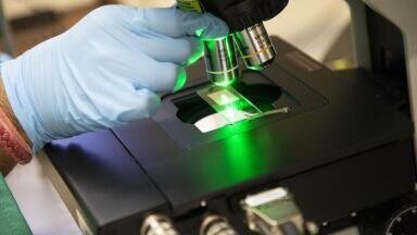
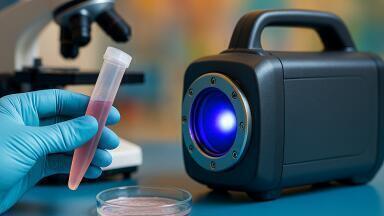
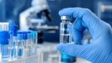
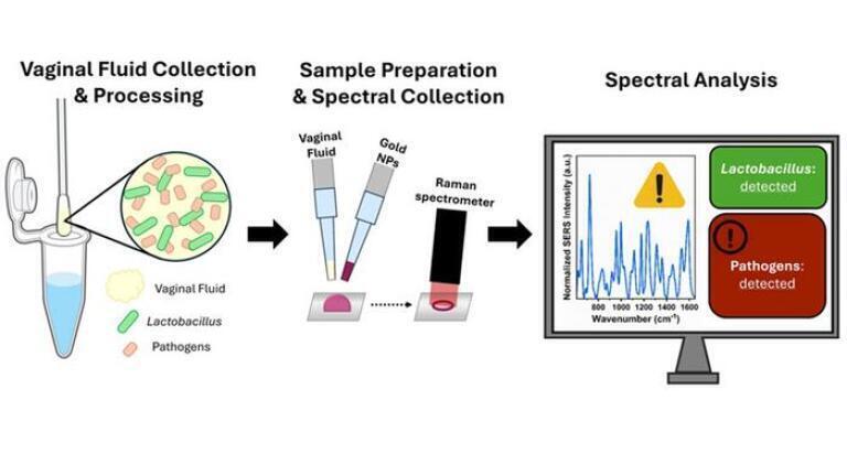
Vaginal health is closely linked to the balance of bacteria within the microbiome, particularly species of Lactobacillus. Disruption of this balance – a condition known as dysbiosis – can increase the risk of infection, pregnancy complications and longer-term health concerns. Current diagnostic methods can often fail to detect Lactobacillus iners, a key bacterium which is difficult to identify under the microscope or in culture. A research team fromVanderbilt University Nashville, Tennessee, has used surface-enhanced Raman spectroscopy (SERS) to analyse the biochemical fingerprints of vaginal fluid in order to address these limitations.
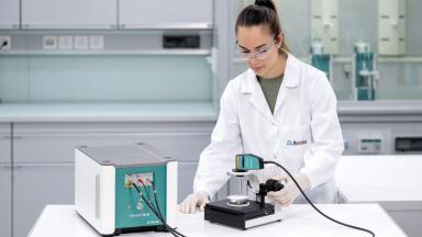
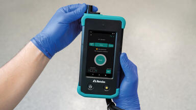
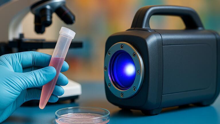
In a pilot study the team collected samples from 19 participants during their routine gynaecological examinations. They used both a laboratory Raman microscope and a portable Raman spectrometer to record SERS spectra, revealing the presence of proteins, lipids, organic acids and sugars. The microbial composition was determined using quantitative polymerase chain reaction (PCR) to identify L. iners, Lactobacillus crispatus, Gardnerella vaginalis and Streptococcus agalactiae.
The researchers found that G. vaginalis – which is associated with bacterial vaginosis – correlated with increased protein and lipid signals and reduced organic acid content, which is consistent with its role in disrupting the vaginal environment.
L. iners showed the opposite trend, with elevated organic acids and reduced protein and polysaccharide signals. These biochemical patterns were visible using both benchtop and portable devices.
Samples containing G. vaginalis were obtained from participants with no diagnosed infection or symptoms, suggesting that SERS could detect subclinical microbiome shifts before they become clinically apparent. The comparable performance of the portable device indicated that accurate biochemical analysis does not require a full laboratory setting.
Although the pilot scheme assessed only a limited number of bacterial species, the authors reported that the work provided proof of concept for the use of SERS in vaginal microbiome assessment. The team plans to expand its participant pool and employ genetic sequencing for broader microbial analysis, with the aim to develop faster, more objective diagnostic tools to improve women’s health.
For further reading please visit: 10.1117/1.BIOS.2.4.042102
ILM 51.5 July 2026


-(1).jpg)
.jpg)