Raman spectroscopy
University at Albany researchers have reported a deep ultraviolet Raman spectroscopy technique to assess messenger RNA encapsulation within lipid nanoparticles, with potential implications for quality control of mRNA therapeutics
Messenger RNA (mRNA) has transformed modern medicine by instructing cells to, among other things, produce proteins that enable the immune system to prevent or combat disease. Its impact has become particularly evident in vaccines and experimental therapeutics that target cancer and rare genetic disorders. Yet the clinical success of such interventions has depended not only on molecular design but also on the stability and delivery of the fragile genetic material itself.
Before administration mRNA requires encapsulation within lipid nanoparticles. These microscopic lipid spheres shield the molecule from rapid enzymatic degradation and facilitate entry into target cells, where the encoded protein can be expressed. If encapsulation fails or remains incomplete, both safety and efficacy could suffer.
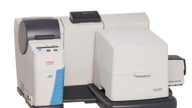
Researchers at the University at Albany, part of the State University of New York system, New York, USA, have now developed a technique to determine whether mRNA sits securely within these lipid carriers. In a recent study the team described a Raman spectroscopy approach that enables rapid, non-destructive analysis of intact samples.
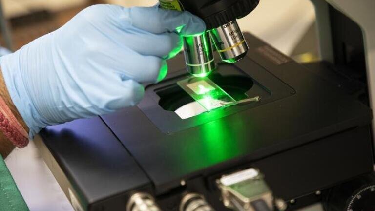
Raman spectroscopy relies on the interaction between laser light and molecular vibrations. When a monochromatic laser illuminates a sample, most light scatters elastically. A small fraction scatters inelastically, with shifts in wavelength that reflect the vibrational modes of the molecules present. Each chemical structure therefore produces a distinctive spectral pattern, often described as a molecular fingerprint.
“mRNA therapeutics have emerged as a powerful tool for treating a wide range of diseases but their clinical success depends on overcoming issues of instability and delivery,” said Dr. Igor Lednev, who is the Williams-Raycheff Endowed Professor in the Department of Chemistry and the RNA Institute, at the University at Albany, and led the work.
“Raman spectroscopy offers us unique information that can help to ensure mRNA is fully encapsulated inside lipid nanoparticles, ensuring the safety and effectiveness of these therapeutics,” he said.
The Raman approach described by the Albany team preserves sample integrity and yields results without lengthy preparation.
“Intact lipid nanoparticles are not very stable and are difficult to characterise by existing techniques,” said Dr. Alexander Shekhtman, professor in the Department of Chemistry at the University at Albany and researcher at the RNA Institute.
“Raman spectroscopy allows us to analyse mRNA inside lipid nanoparticles without damaging it. This means we can optimise formulations to improve both safety and effectiveness,” he added.
Detecting mRNA within a lipid nanoparticle presents a technical challenge because the nucleic acid constitutes only a small fraction of the total mass. The surrounding lipids dominate the chemical signal. To address this limitation, the researchers have employed a specialised deep ultraviolet Raman instrument constructed within the Lednev laboratory. Deep ultraviolet excitation enhances the signal from nucleic acids while it reduces interference from lipid components which absorb less strongly in that wavelength range of the spectrum.
“By using our homebuilt instrument to directly analyse mRNA molecules in vaccine samples – and by combining this with advanced statistical analysis – we have created a quantitative method to ensure that the mRNA is properly protected within lipid nanoparticles,” Lednev said.
The team has integrated machine learning algorithms with spectroscopic data to extract subtle molecular signatures from complex mixtures.
Over the past two decades, Lednev has pioneered the application of Raman spectroscopy in forensic science and medical diagnostics. His previous work has included methods to identify biological stains, gunshot residue, hair and trace evidence at crime scenes, as well as approaches to enable non-invasive early diagnosis of neurodegenerative conditions such as Alzheimer’s disease.
The researchers believe that the present method could serve in quality control environments to evaluate mRNA therapeutics before release and to support formulation development during research and development. As mRNA platforms expand the need to verify structural integrity will become more pressing.
“This is an example of how advances in [Raman] laser spectroscopy can directly support modern medicine. By better understanding how these therapeutics are formulated, we can help to make them safer and more effective,” Lednev said.
For further reading please visit: 10.1021/acs.analchem.5c04246
ILM 51.5 July 2026


-(1).jpg)
.jpg)