.jpg)
Laboratory products
Published over 5 years ago. See the latest and most current information on Laboratory products.
Image analysis is one technique of particle characterisation which can provide many tangible benefits in comparison to more traditional methods. For example, it can contribute greater informational content and increased accuracy, while it is unique in its ability to deliver reliable data about the shape parameters of the particles in question, including their aspect ratio and roundness.
In recent years, image analysis has become an ever more popular technique of particle characterisation, which has led to an increased amount of research into developing the capabilities of the medium. Generally speaking, there is a broad distinction between static and dynamic analyses, both of which fields have seen impressive advances in the last few years. Here’s a quick look into the particulars of those advances as they pertain to the field of particle characterisation.
State image analysis employs a microscopic procedure which takes photographs of a sample slide at each phase of the experiment, then automatically evaluates those images. It is appropriate for samples with narrow size distributions where it is desirable to obtain more accurate and precise information about finer particles.
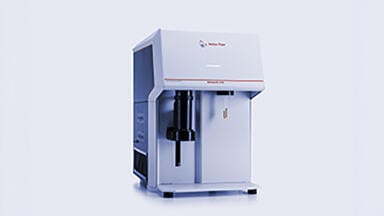
One of the most exciting recent developments in this field is the release of the CAMSIZER M1, which uses five lenses to achieve a comprehensive measuring range of between 0.5µm and 1,500µm. The device automatically analyses the particle size and shape of the sample and determines which focal range is most appropriate, ensuring optimum results every time. For those interested, the article How to perform fully automated static image analysis simply and accurately contains more information about this innovative new development.
As the name suggests, dynamic image analysis differs from its static counterparts in that it captures images of particles while they are in motion. This offers researchers greater insight into how the particles behave for the duration of the experiment and the ability to determine not only if a specific process has occurred, but also pinpoint the moment at which it began and the speed with which it unfolded.
The CytoSMART Lux3 FL is one of the most impressive new technologies in this area of research. A portable, live-cell imaging microscope whose small size does not seem congruent with the myriad features and benefits it carries, the device is equipped with one brightfield and two fluorescent channels, one of which is red and the other green. This gives users the option to track molecular processes in real-time in a laboratory-controlled setting. For more information on the technology, check out the article New Fluorescence Live-cell Imaging Microscope Launched.
ILM 51.5 July 2026


-(1).jpg)
.jpg)

.jpg)