Research news
Researchers at Baylor College of Medicine have reported a novel blood-based method to detect circulating tumour cells in triple negative breast cancer, identifying four surface proteins that enable more sensitive monitoring of metastatic disease
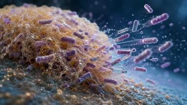
Triple negative breast cancer is widely recognised as the most aggressive form of the disease and remains without targeted therapies. It is also more likely than other breast cancer subtypes to metastasise, with tumour cells travelling around the body’s bloodstream. This process accounts for more than half of breast cancer-related deaths each year. Despite the clinical importance of metastasis, it has remained difficult to track reliably because circulating tumour cells are rare and lack distinctive markers that clearly separate them from normal blood cells.
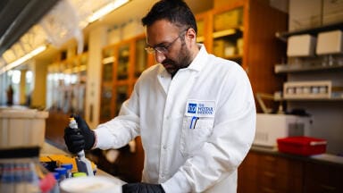
Researchers at Baylor College of Medicine, Houston, Texas, USA, have now developed a procedure to enhance the detection of circulating tumour cells in triple negative breast cancer using a simple blood draw, often described as a ‘liquid biopsy’. The approach offers a minimally invasive way to monitor cancer progression in near real time and has led to the identification of four proteins expressed on the surface of live circulating tumour cells that specifically distinguish these cells from normal blood components.
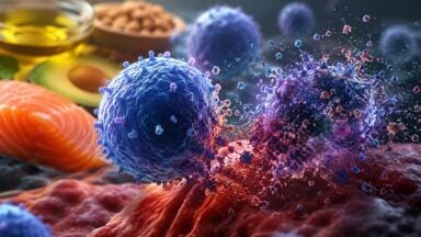
The ability to capture live circulating tumour cells is particularly important because it allows researchers to analyse the genetic material of individual cells. This level of resolution supports detailed investigation of how cancer spreads, how tumour cells evolve during disease progression, and why some cancers fail to respond to treatment.
“We developed a workflow [which can] isolate and analyse live circulating tumour cells, focusing first on mouse models of metastatic triple negative breast cancer and then testing our findings in patient samples,” said first author Dr Bree M. Lege, formerly a graduate student in the laboratory of corresponding author Dr Chonghui Cheng.
Dr Cheng is a professor of molecular and human genetics and of molecular and cellular biology and is based in the Lester and Sue Smith Breast Center at Baylor. She is also a member of the Dan L Duncan Comprehensive Cancer Center.
The research team began by capturing live circulating tumour cells from the blood of tumour-bearing mice. Because these cells are extremely scarce, the researchers first separated tumour cells from normal blood cells. Individual tumour cells were then isolated and analysed using single-cell RNA sequencing, a technique that measures gene activity within each cell. This analysis enabled the team to identify which genes were active and – critically – which proteins were present on the surface of circulating tumour cells in triple negative breast cancer.
Through this approach, the researchers identified four surface markers, AHNAK2, CAVIN1, ODR4 and TRIML2, that were present on circulating tumour cells but absent from normal blood cells. The lack of overlap between these markers and proteins expressed by healthy blood cells reduces the likelihood of false positive results, a longstanding challenge in liquid biopsy technologies.
“We are very pleased with our approach to identify circulating tumour cells in blood,” Cheng said.
“The novel markers detected cells that standard methods missed. When the four markers were combined, detection improved substantially.
“Importantly, the markers showed very little overlap with those found on normal blood cells, which reduced the risk of false positives,” she added.
The researchers then applied the same strategy to blood samples from patients with metastatic triple negative breast cancer. In these samples, tumour cells were often undetectable using conventional markers but became clearly visible when the newly identified marker combination was applied.
“We were excited by the results obtained from blood samples from patients with metastatic triple negative breast cancer,” Cheng said.
“In these patients, tumour cells frequently evaded detection with standard markers but became readily detectable with our marker panel,” she concluded.
The findings address a major limitation in current liquid biopsy approaches for aggressive breast cancers. Reliable detection of circulating tumour cells in triple negative breast cancer could allow clinicians to monitor disease progression and treatment response with greater accuracy and sensitivity. Because the method enables capture of live cells, it also opens the way for detailed analysis of tumour cell gene expression, which may help to explain how metastasis occurs and why resistance to therapy develops.
Beyond breast cancer, the researchers also reported that the newly identified markers are expressed in other cancer types. This observation suggests that the same strategy could improve circulating tumour cell detection across a broader range of malignancies, with potential implications for cancer monitoring and personalised treatment strategies.
For further reading please visit: 10.1158/2767-9764.CRC-25-0536
ILM 51.5 July 2026


.jpg)
.jpg)


-(1).jpg)