Research news
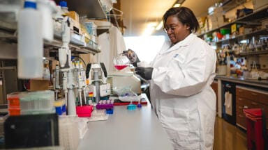
Scientists at National Taiwan University (NTU) have developed a pioneering optical microscopy method that can reconstruct a person’s long-term blood glucose history from a simple blood sample, opening new frontiers for diabetes and cancer research.
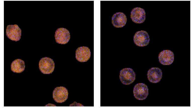
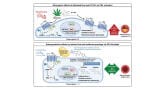
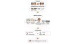
The interdisciplinary team, led by Professors Chi-Kuang Sun and Tzung-Dau Wang, developed colour-resolved third-harmonic-generation microscopy (cTHGM) - a label-free imaging technique capable of distinguishing glycated haemoglobin (HbA1c) from normal haemoglobin inside individual red blood cells.
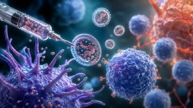
Unlike current monitoring systems, which capture only short-term glucose levels, the new approach allows researchers to trace blood sugar fluctuations over the 120-day lifespan of red blood cells. This effectively provides a ‘molecular time-lapse’ of a person’s metabolic history.
To achieve this, the NTU team built a broadband, femtosecond-laser-based microscope that detects nanometre-scale wavelength shifts caused by chemical differences in haemoglobin molecules. The result is a non-invasive, high-resolution chemical map showing how much glucose each red blood cell has encountered during its lifetime.
“If continuous glucose monitoring gives you a short clip of daily sugar changes, our imaging method offers the full 120-day documentary,” explained Professor Sun.
The researchers believe cTHGM could enhance precision diabetes management and help identify early signs of metabolic disorders linked to cancer. Beyond clinical use, the technique opens possibilities for real-time, colour-sensitive molecular imaging in living tissues.
The study [1], published in Science Advances, demonstrates how advanced microscopy can turn invisible biochemical traces into powerful diagnostic information.
More information online
ILM Guide 2026/27


.jpg)


2.jpg)
