Research news
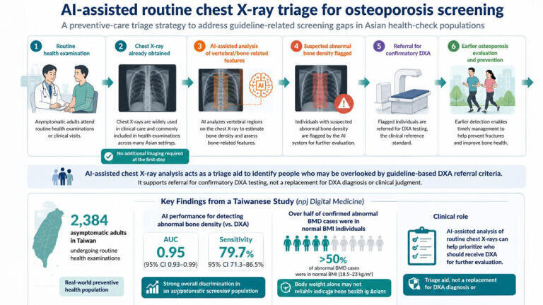
Researchers from National Taiwan University and St. Paul’s Hospital, Taiwan have shown that artificial intelligence can extract signs of asymptomatic bone loss from routine chest X-rays, offering a potential route to expand osteoporosis screening beyond current clinical guidelines.
Osteoporosis is often described as a ‘silent disease’, developing gradually over time before fractures occur. Existing screening recommendations largely focus on older women and selected high-risk groups, meaning some men, younger adults, and individuals with normal body weight are not routinely assessed.
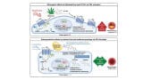
To address this gap, the research team demonstrated in npj Digital Medicine [1] that AI models can identify indicators of reduced bone density using chest X-rays that are already widely taken during routine health checks.
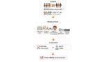
A notable finding from the study was that more than half of confirmed cases of low bone density occurred in individuals with a normal body mass index (BMI), highlighting a potential blind spot in current screening approaches based on risk profiles alone.
Because chest X-rays are already routinely collected in many healthcare systems, particularly in Asia, the approach offers a low-cost, infrastructure-light opportunity for opportunistic screening without additional patient burden. The system could help flag individuals outside traditional risk groups who may benefit from follow-up dual-energy X-ray absorptiometry (DXA) testing.
“Under Taiwan’s National Health Insurance system, we often rely on strict guideline-based criteria to decide who qualifies for DXA testing,” said Shu-Han Chen, MD, first author of the study and a physician at St. Paul’s Hospital. “Our findings suggest that AI-assisted chest X-ray analysis could help identify individuals who may otherwise be overlooked and who may benefit from confirmatory testing.”
“This study demonstrates how artificial intelligence can transform existing healthcare workflows into scalable preventive strategies, while supporting more equitable access to osteoporosis screening,” added Professor. Ray-E Chang from National Taiwan University.
The researchers suggest that AI-assisted interpretation of routinely acquired imaging could help bridge gaps in current screening pathways, enabling earlier identification of at-risk individuals who might otherwise remain undiagnosed until fracture occurs.
More information online
Lab Asia 33.4 - August 2026


-(1).jpg)


-(1).jpg)
