News
Published over 10 years ago. See the latest and most current information on News.
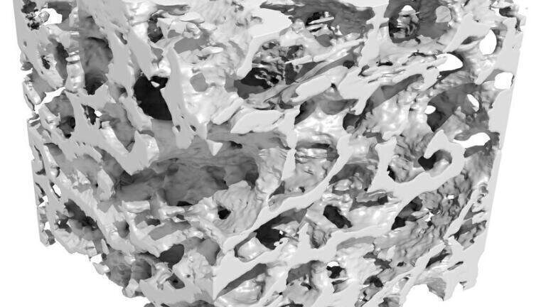
A new method of taking highly detailed x-ray images of bone using powerful laser beams has been developed by researchers at Imperial College London (ICL) in collaboration with the lasers team at the Science and Technology Facilities Council (STFC). The x-rays were produced using a laser wakefield accelerator – a compact type of particle accelerator developed at ICL- to produce three-dimensional images of bone samples at resolutions of around 50 microns. This would allow clinicians to spot features within the bone that were finer than a human hair – at around 100 microns thick.
Following early stage results the scientists are now working on refining their technique and exploring how the technology could be adapted for use in a hospital. In particular, work is underway to also miniaturise the laser system that underpins this work so that the whole system can be made to fit into a hospital imaging department.
“Wakefield accelerators have been developed with the idea that they could make the accelerators used in high-energy physics experiments, such as at CERN, much more compact and thus cheaper. However, those same benefits could see wakefield accelerators being used in other applications of accelerators, such as in generating high quality beams of x-rays” commented research team leader Professor Zulfikar Najmudin, of the John Adams Institute for Accelerator Science in the Physics Department at Imperial.
The team have been developing their technique at STFC’s Central Laser Facility, based at the Rutherford Appleton Laboratory in Oxfordshire.
“This demonstration of high resolution x-ray imaging builds on many years of research conducted using our world-leading systems – in this case the Gemini laser – and brings together experienced researchers from different fields”, said STFC’s Dr Dan Symes, one of the research team.
“In this study, we’ve shown that the laser technology has the potential to be adapted so that we can create x-rays that reveal unprecedented detail,” explains Jason Cole, lead author of the study, also from Imperial. “This kind of high-resolution x-ray would be extremely useful for identifying conditions such as osteoporosis. Conventional x-ray scanning cannot get the full picture, as early-stage bone cracks can be too fine to be picked up.”
The team used this beam to produce pictures of a small sample from the leg bone taken from an elderly patient who had had a hip replacement. By rotating the bone slightly after each picture, a full three-dimensional reconstruction of the sample was produced showing fine detail of the bone’s internal structure.
“The internal structure of bone is surprisingly complicated – you need powerful x-rays to penetrate the material deeply enough to get a detailed image,” said Cole. “Our method was able to produce really detailed pictures that could be transformed into a 3D model of the sample.”
The team expects that, with refinement, their system could provide a practical alternative to microtomography.
The research was funded by the Engineering and Physical Sciences Research Council and the Science and Technology Facilities Council in the UK.
Published in Scientific Reports, Aug 18, 2015
ILM Guide 2026/27


.jpg)