Chromatography
Published over 6 years ago. See the latest and most current information on Chromatography.
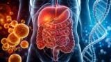
In a scientific breakthrough that could revolutionise cardiology, a team of researchers from Carnegie Mellon University in Pittsburgh has developed a 3D printer that uses collagen to manually reconstruct components of the human heart and build complex collagen scaffolds that replicate the look, feel and function of biological tissues.
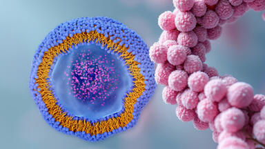
As the main component of most tissues and organs in the human body, collagen is an incredibly versatile protein. Until now, it's been difficult to print living cells and soft biomaterials such as collagen because of the unstable nature. The new 3D printer developed by the Carnegie Mellon University overcomes this challenge with the use of a support gel. The gel allows the printer to create collagen-rich heart tissue and build parts of the organ, including capillaries, chambers, valves and vessels.
The technique is known as FRESH, short for freeform reversible embedding of suspended hydrogels. A report published in the journal Science explains how the team overcame the challenge of 3D printing "soft and squishy" tissues by using a stabilising gel. "In our work we have solved this problem by 3D bioprinting inside a ‘support’ gel that prevents the cells and hydrogels (similar to gelatine) from collapsing,” explains study leader Adam Feinberg.
Armed with the new technique, Feinberg and his team were able to successfully 3D print collagen with the same ease as other materials such as metal and plastic. “Being able to do this means that we can now print scaffolds and tissues that actually have biological function,” he asserts.
The method works by adding a supply of supportive gelatine microparticle slurry to the 3D printer. Bioinks containing unmodified collagen are then inserted into the printing needle, which creates thin, stacked layers of collagen and cells. After the printing process is complete, the room temperature is raised to 37°C which slowly melts the support gel and releases the printed collagen scaffold.
As well as general heart components, Feinberg also says FRESH can be used to print patient-specific anatomical structures using MRI or CT images. The images can then be used to generate unique 3D computer models and printing instructions that meet the exact needs of the patient.
If you like the concept of FRESH, you'll also find the revolutionary new LSER method fascinating. Find out more about how scientists are building an understanding of the interactions between analyte and sorbent by reading 'Characterisation of RP Sorbents by Linear Solvation Energy Relationships (LSER).'
ILM 51.5 July 2026


.jpg)
.jpg)


-(1).jpg)