Clinical
Scripps Research team offers insights that could aid development of phage therapies against Mycobacteria
The pathogens which cause infectious diseases including tuberculosis (TB) – Mycobacteria – are the world’s most deadly bacteria, responsible for more than one million deaths annually. Antibiotic-resistant mycobacteria is on the rise meaning new drugs to fight these infections are urgently needed.
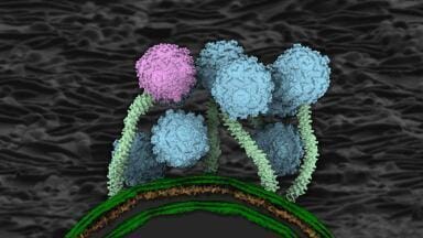
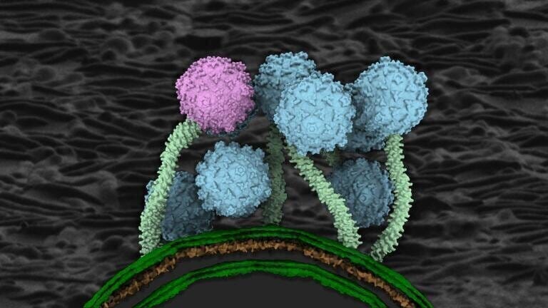
A research team from Scripps Research and the University of Pittsburgh has now imaged a type of virus – known as a bacteriophage, or just ‘phage’ – which invades Mycobacteria. The research could lead to phage-based treatments for mycobacteria that are resistant to current antibiotics.
“Phages have evolved over millions of years to precisely target specific bacteria. But to be able to develop phages into effective therapies, we need to know more about how they interact with Mycobacteria,” said Assistant Professor Donghyun Raphael Park at Scripps Research, and a co-senior author of the article.
Phage therapies – using viruses to attack drug-resistant bacteria – are being examined as alternatives to typical antibiotics. The phages can recognise different aspects of bacteria when compared to antibiotics and so may be able to kill bacterial pathogens that have become drug resistant.
However, the phages that target Mycobacteria – known as mycobacteriophages – are poorly understood in terms of their structures or how they infect Mycobacteria.
Park’s colleagues included Professor Graham Hatfull of the University of Pittsburgh and Howard Hughes Medical Institute, who’s expertise assisted in creating atomic-level models of a mycobacteriophage known as Bxb1.
They captured images at multiple stages of infection – revealing how Bxb1 attaches to Mycobacteria, inserts its own genetic material. Combining data from a single particle cryo-electron microscopy (cryo-EM) and cryo-electron tomography (cryo-ET), allowed the research team to use the two imaging techniques together to visualise frozen biological structures at near-atomic resolution.
“Other phages form a channel through the bacterial membrane to inject their DNA. We expected to see the same here,” Park said.
“But we didn’t. This suggests mycobacteriophages use a completely different genome translocation mechanism.”
Compared to other bacteria, Myobacteria have unusually thick cell walls and more work is needed to understand how phages can insert their genome through this formidable cell wall defence.
Park said that detailing the structures of other mycobacteriophages should reveal what structural elements – such as this tail tip – are most important. For example, the imaging revealed how the tail tip of the phage dramatically changed when it bound to the bacteria, suggesting this as a dynamic process of infection.
“There are thousands of mycobacteriophages out there, but we don’t yet fully understand how they recognise and kill Mycobacteria,” Park said.
“By continuing to study their structures, we can start to identify the hallmarks of an effective phage and design better treatments.”
While Park doesn’t plan to work out the structures of all of the thousands of phages that could fight Mycobacteria, his lab will focus on a few more and delve into studies linking the phages’ structures to their functions. This could guide the rational selection of phages for treating Mycobacteria, helping other researchers to identify which phages may work best for the design of an effective phage therapy to antibiotic-resistant TB.
In addition to Park and Hatfull, authors of the study, “Structure and infection dynamics of mycobacteriophage Bxb1,” were Sudipta Mondal, Benjamin Silva and Valery Ortiz of Scripps; Krista Freeman, Deborah Jacobs-Sera, Alexis Huet, and James Conway of University of Pittsburgh; Lourriel Macale, Ronelito Perez, Joemark Narsico, Meng-Chiao Ho, and Todd Lowary of Academia Sinica; Jennifer Podgorski and Simon White of University of Connecticut.
For further reading please visit Science Direct
ILM 51.5 July 2026


-(1).jpg)
.jpg)