Clinical
Non-destructive micron-resolution imaging of cochlea lays groundwork for better hearing loss diagnosis
For the first time, researchers have shown that terahertz imaging can be used to visualise internal details of the mouse cochlea with micron-level spatial resolution. The non-invasive method could open new possibilities for diagnosing hearing loss and other ear-related conditions.
A multi-institutional group of researchers have developed a 3D terahertz near-field imaging technique, which provides high-resolution images that can be used to create detailed 3D reconstructions.
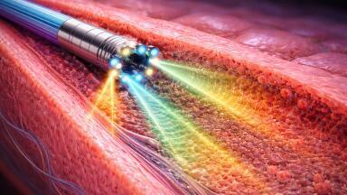
Terahertz radiation, which falls between microwaves and the mid-infrared region of the electromagnetic spectrum, is ideal for biological imaging because it is low-energy and non-harmful to tissues. It scatters less than near infrared and visible light and can pass through bone while also being sensitive to changes in hydration and cellular structure.
“Hearing relies on the cochlea, a spiral-shaped organ in the inner ear that converts sound waves into neural signals,” said Associate Professor Kazunori Serita, and research team leader, from Waseda University in Japan.
“Conventional imaging methods often struggle to visualise this organ’s fine details. Our 3D terahertz near-field imaging technique allows [for] small structures inside the cochlea [to be seen] without any damage,” said Serita.
“This technique could lead to a new [examination] method for ear diseases that have been difficult to diagnose until now. It has the potential to enable on-site diagnosis of conditions like sensorineural hearing loss [as well as] other ear disorders.
“It might also be useful for early detection of hearing impairments, allowing for earlier treatment and better outcomes,” he added.
Serita was inspired to develop the technique after learning about cochlear measurement challenges from coauthor Associate Professor Takeshi Fujita from the Department of Otolaryngology-Head and Neck Surgery at Kobe University.
“That got me thinking – maybe terahertz imaging could help solve these issues,” said Serita.
“We decided to collaborate. The big question was whether we could visualise the tiny internal structures of the cochlea without causing any damage.”
Terahertz imaging is typically performed by focusing terahertz waves using a lens made for these wavelengths. However, these lenses are typically limited to focal sizes measuring a few millimetres – too large to image the tiny structures of the cochlea.
In this work, the researchers eliminated the need for a focal lens by using a nonlinear optical crystal to create terahertz light emitting from a very small region within the crystal. Because this terahertz point source had a beam diameter of just 20 microns, the researchers could measure much smaller samples with terahertz waves.
"Until now, there was no way to observe the internal structure of the cochlea non-destructively with high resolution,” said Serita.
“A key innovation in our work was the use of a nonlinear optical crystal to generate terahertz waves from 1,560-nm near-infrared light. This was crucial for our imaging technique.”
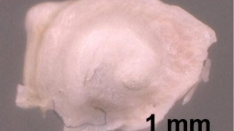
Testing their approach, the researchers needed to confirm that the terahertz waves were reaching the inside of the cochlea. They did this by using the terahertz imaging setup to conduct experiments using two different extracted and dried mouse cochlear samples – one with an empty interior and another filled with a metal material that reflects terahertz waves. They observed clear differences between the two samples, confirming that the terahertz waves were penetrating the inside of the cochlea.
The researchers then showed that internal structural information could be easily observed and extracted from 2D terahertz time-domain images using an unsupervised learning algorithm. The team also used the setup to successfully carry out 3D terahertz time-of-flight imaging and 3D reconstruction, allowing visualisation of part of the cochlear duct – the spiral structure inside the cochlea.
Next, the researchers plan to demonstrate the technique’s feasibility on cochleae in a more realistic biological environment. Since the cochlea is located deep inside the ear and filled with lymphatic fluid, they will first need to miniaturise the system so it can be inserted through the ear canal. They are also developing a stronger terahertz source to reach deeper structures.
The researchers say that once the terahertz imaging technology is miniaturised, it could be incorporated into endoscopes and otoscopes, enabling non-invasive in vivo imaging for cochlear diagnostics or early cancer detection in various organs.
For further reading please visit: 10.1364/OPTICA.543436
ILM 51.5 July 2026


-(1).jpg)
.jpg)