Clinical
A compact side-viewing fibre probe for endoscopic optical coherence tomography has achieved micrometre-scale resolution across an unprecedented imaging depth, addressing a long-standing trade-off that has limited minimally invasive diagnosis in narrow anatomical spaces
Seeing fine tissue structures deep within the body has long required compromise. High-resolution optical images typically degraded rapidly with depth, while attempts to visualise deeper regions resulted in a loss of sharpness. Researchers have now reported a miniature side-viewing fibre probe that has overcome this constraint, enabling clinicians to image deeper tissue while preserving fine structural detail.
The probe remains extremely small, which makes it suitable for narrow and sensitive anatomical environments, and combines extended imaging depth with micrometre-scale resolution to provide a clearer and more reliable view of internal structures. This capability has the potential to improve the detection of early-stage tissue abnormalities during minimally invasive procedures.
Endoscopic optical coherence tomography is widely used to visualise tissue microstructures in real time but existing probe designs have faced persistent limitations. Conventional probes struggle within narrow lumens where space is restricted and the risk of tissue damage must be minimised. A fundamental physical trade-off has also constrained performance, since attempts to increase lateral resolution have reduced imaging depth, while efforts to extend depth have blurred fine details.
These factors have limited the clinical value of endoscopic imaging, particularly for early diagnosis in confined structures. Difficulties associated with probe manufacture have further restricted miniaturisation and robustness, underlining the need to develop side-viewing probes that can deliver both deep and sharp imaging without increases in size or complexity.
A research team led by scientists at Beijing Institute of Technology, China, has presented a side-viewing fibre probe for optical coherence tomography that addressed these challenges. The probe introduced a redesigned strategy for light delivery that substantially extended imaging depth while preserving high lateral resolution. Tests that used both linear and rotational scanning produced clear images in biological tissues and narrow-lumen samples, indicating a practical route towards safer and more informative endoscopic imaging in clinical and industrial contexts.
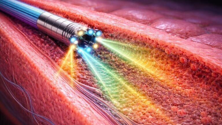
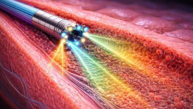
The advance centred on how the probe controlled light propagation. Rather than focusing light into a single tight spot that rapidly diverged, the probe maintained a narrow beam over an extended distance. This approach allowed the system to capture sharp images across a much larger depth range than had previously been possible with fibre-based probes.
Experimental results showed that the probe achieved an imaging depth of approximately 350 micrometres, which was more than ten times greater than that of many conventional fibre probes, while maintaining a lateral resolution of around 1.4 micrometres. In practical terms, fine structural features remained visible even as imaging extended deeper into tissue.
Crucially, this performance was delivered in a probe with a diameter close to one millimetre, small enough to navigate narrow anatomical passages. Imaging quality also remained stable during rotational scanning, which is essential for three-dimensional endoscopic imaging.
The probe successfully resolved internal features within layered materials, plant tissues, and animal tissues. These demonstrations showed that extended imaging depth and high resolution no longer need to be traded against one another, but can instead coexist within a compact, fibre-based design suitable for real-world use.
“This work shows that we can rethink the limits of miniature endoscopic imaging,” said the study’s corresponding author, Dr. Yong Huang.
“By keeping the beam focused over a longer range, we can see deeper while preserving fine detail. Just as importantly, the probe is built using standard fibre-processing techniques, which makes it realistic to scale and deploy.
“We believe this approach can help bring more reliable, less invasive imaging tools into clinical practice,” Huang added.
The fibre probe could broaden the application of endoscopic optical coherence tomography in areas where imaging has previously been difficult or risky. In medicine, clearer visualisation of airways, gastrointestinal tracts, and paediatric organs could support earlier diagnosis with reduced tissue disturbance.
Beyond healthcare, the technology could be adapted for non-destructive inspection of industrial components, layered materials, or microscale defects. Because the design is compact, cost-effective, and compatible with existing manufacturing methods, it offers a realistic pathway from laboratory research to deployable devices. More broadly, the study illustrates how improved control of light propagation can redefine the capabilities of miniature imaging systems.
For further reading please visit: 10.1038/s41378-025-01034-x
Lab Asia 33.4 - August 2026


-(1).jpg)


-(1).jpg)
