Research news
Researchers have identified a lipid-driven biophysical mechanism that allows tuberculosis-causing mycobacteria to survive inside human immune cells, a discovery that could inform novel therapeutic strategies
Scientists have uncovered an elegant biophysical mechanism that tuberculosis (TB)-causing bacteria use to survive within human immune cells, a finding that has offered a fresh perspective on how the pathogen evades destruction and persists in the body. The work has suggested that subtle changes to membrane physics – rather than protein interference alone – may underpin the bacterium’s enduring and deadly success.
TB, caused by members of the genus Mycobacterium, has remained the world’s leading infectious killer. The disease claims more than a million lives each year and continues to pose a major public health challenge across Asia, Africa and Latin America. Despite decades of research, the bacterium’s ability to subvert host immunity has frustrated all eradication efforts.
“TB is rampant in India,” said doctoral candidate Ayush Panda, formerly a graduate student in the laboratory of Mohammed Saleem with the School of Biological Sciences, National Institute of Science Education and Research, Bhubaneswar, India.
“I grew up in a (Indian) state where TB outbreaks are a major problem and I was always curious about how these diseases spread [drawing] me to this research,” he added.
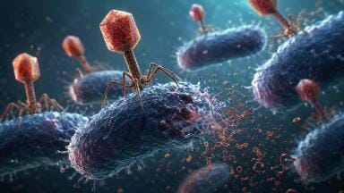
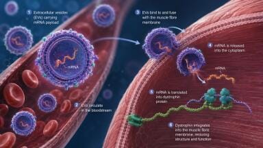
The team focused on extracellular vesicles, which are nanoscale membrane-bound particles that cells release to transport molecular cargo. The researchers found that mycobacteria released extracellular vesicles that fused with the membranes of host immune cells. These vesicles carried specialised lipids – fatty molecules with structural and signalling roles – that altered the physical properties of the host membrane.
Under normal circumstances, immune cells such as macrophages engulf invading bacteria and enclose them within membrane-bound compartments known as phagosomes. The phagosome then fuses with a lysosome, an acidic, enzyme-rich organelle that digests and destroys microbial invaders. In this process the fusion step is essential for effective bacterial clearance.
The researchers discovered that mycobacterial lipids stiffened the phagosome membrane. Increased membrane rigidity reduced its capacity to merge with the lysosome. As a result, the bacterium remained protected within a compartment that failed to mature into a destructive environment. In effect, the pathogen constructed a protective niche inside the very cell that was seeking to eliminate it.
“If the membrane becomes more rigid, it becomes much harder for the phagosome to fuse with the lysosome.
“It is an elegant biophysical mechanism – the bacteria remodelled the membrane architecture to escape the very process that would have killed them,” Panda explained.
The study also demonstrated that these extracellular vesicles did not confine their effects to infected cells. Vesicles could diffuse to neighbouring immune cells and modify their membranes before direct bacterial contact occurred. This observation suggested that mycobacteria may prepare a permissive environment in advance of infection, thereby dampening host defences at a tissue level.
What set this work apart was its lipid-centric focus. With much previous research into TB pathogenesis having concentrated on bacterial proteins that interfere with host signalling pathways. By contrast, this study examined the physical consequences of lipid transfer. The researchers introduced purified mycobacterial lipids into artificial membranes designed to mimic the phagosome and observed marked changes in membrane properties.
“The most surprising finding was when we introduced mycobacterial lipids into membranes that mimic the host phagosome, we saw remarkable physical changes – the membrane properties were completely altered,” Panda said.
These experiments have indicated that lipid insertion alone was sufficient to induce mechanical changes that impair immune function. Such findings have broadened the conceptual framework through which scientists understand host – pathogen interactions. They have highlighted that infection is not only a biochemical contest but also a physical one, in which membrane tension, curvature and rigidity shape cellular outcomes.
The team extended its observations to other bacterial species with similar extracellular vesicle-mediated effects on membrane stiffness being observed in the gram-negative Klebsiella pneumoniae, which sits at the top of the World Health Organization’s 2024 bacterial priority pathogens list, and Staphylococcus aureus, the pathogen behind notable and widespread Methicillin-resistant Staphylococcus aureus (MRSA). This suggested that manipulation of host membrane mechanics may represent an evolutionarily conserved strategy among diverse pathogens.
If bacterial survival depends in part on vesicle production and lipid delivery, then drugs that inhibit vesicle formation or neutralise membrane-stiffening lipids could restore effective phagosome – lysosome fusion. Such approaches could complement traditional antibiotics which often target bacterial growth rather than immune evasion.
“Now that we understand how the bacteria protect themselves, we can start to look for ways to stop them.
“If we can block the bacteria from stiffening those membranes, our immune cells might be able to do their job and stop the infection,” Panda said.
TB research has long emphasised antimicrobial resistance and vaccine development. This study has underscored that the physical state of cellular membranes may prove equally important. As global health systems continue to confront more than a million TB deaths each year, insights that integrate biophysics with microbiology may open novel routes to intervention and strengthen the long-standing effort to control this disease.
For further reading please visit: 10.64898/2025.12.17.694930
ILM 51.5 July 2026


.jpg)


-(1).jpg)