Research news
A pioneering scanner developed by scientists at the University of Aberdeen could transform breast cancer diagnosis and treatment, potentially reducing the need for repeat surgeries and enabling more personalised care.
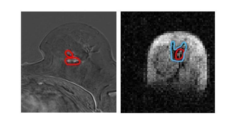
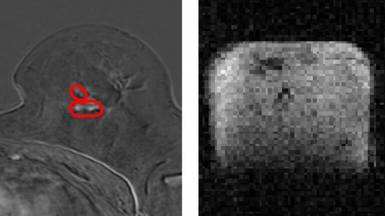
Researchers from the University, in collaboration with NHS Grampian, used a prototype Field Cycling Imager (FCI) to examine breast tissue from newly diagnosed cancer patients. The FCI scanner demonstrated a higher accuracy in distinguishing tumour material from healthy tissue compared to conventional MRI methods.
This breakthrough has the potential to improve treatment outcomes for millions of patients. Currently, around 15% of women require a second surgery after a lumpectomy due to residual tumour cells. By providing more precise tumour mapping, FCI could help reduce the need for additional operations.
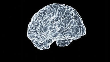
The success of FCI in breast cancer detection follows earlier promising results in identifying brain damage caused by stroke. Developed at the University of Aberdeen, FCI builds on MRI technology but operates at ultra-low magnetic fields, allowing it to detect disease-related changes in organs that were previously impossible to see.
Unlike conventional MRI, which uses strong magnetic fields to generate images, FCI can dynamically adjust magnetic field strength during a scan. This unique capability allows it to extract multiple layers of information from tissue, effectively functioning as multiple scanners in one. Additionally, FCI can detect tumours without the need for contrast agents, which are associated with potential kidney damage and allergic reactions in some patients.
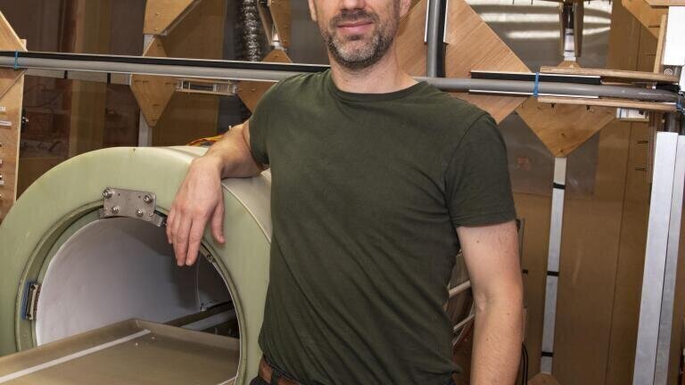
Dr Lionel Broche, Senior Research Fellow in Biomedical Physics and lead researcher, highlighted the significance of these findings: “We found that FCI generates images that characterise breast tumours with greater accuracy. This could enhance biopsy precision, improve treatment planning, and reduce the need for repeat surgeries - offering significant benefits for patients.”
He added: “The University of Aberdeen pioneered the world’s first clinical MRI scanner in the 1970s, and it’s exciting to continue that legacy with an entirely new approach to imaging. As we refine FCI technology, its potential clinical applications are vast.”
Dr Gerald Lip, Consultant Radiologist at NHS Grampian and co-investigator in the study, recently appointed President of the British Society of Breast Radiology, commented: “These early results are promising, and further studies will help validate clinical applications. NHS Grampian treats 400 to 500 women with breast cancer annually, and the potential for FCI to reduce the need for additional surgeries could greatly benefit patients while improving resource efficiency.”
More information online
Published in Nature Communications Medicine
ILM 51.5 July 2026


-(1).jpg)
.jpg)